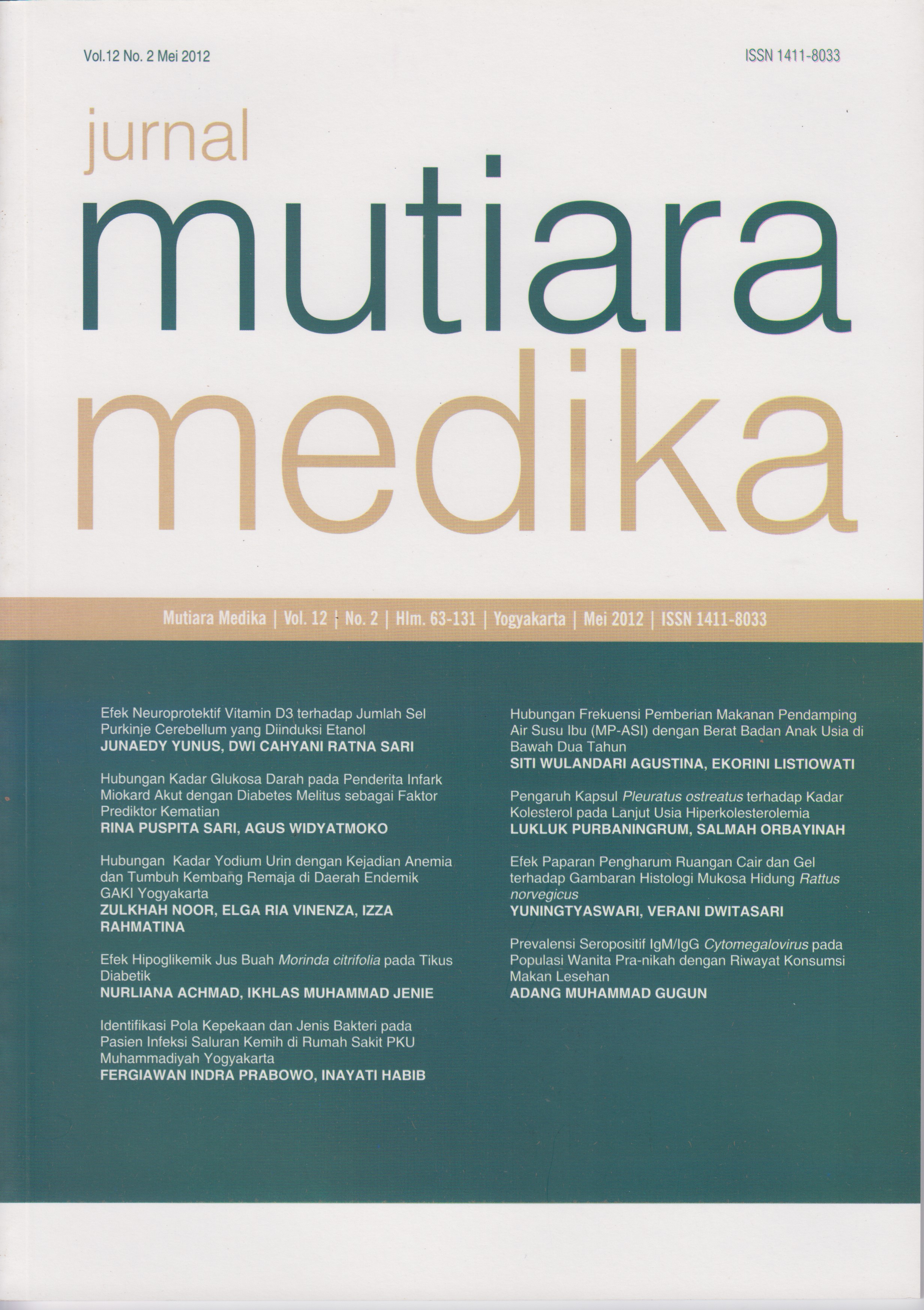
Efek Paparan Pengharum Ruangan Cair dan Gel terhadap Gambaran Histologi Mukosa Hidung Rattus norvegicus
DOI:
https://doi.org/10.18196/mmjkk.v12i2.1023Keywords:
gambaran histologi, mukosa hidung, pengharum ruangan, inhalasi, zat kimia, histologic appearance, nasal mucosa, air freshener, inhalation, chemical substanceAbstract
Pengharum berbentuk cair dan gel merupakan pengharum ruangan modern yang banyak digunakan masyarakat. Berbagai macam zat kimia diduga terkandung di dalamnya, seperti etanol, formaldehida, naftalena, fenol, ptalat dan xilena. Zat kimia pengharum masuk pertama kali ke dalam tubuh melalui rongga hidung. Penelitian ini bertujuan mengkaji perbedaan pengaruh paparan pengharum ruangan cair dan gel terhadap gambaran histologi mukosa respiratorius rongga hidung. Jenis penelitian adalah eksperimental dengan post-test only control group design. Subyek penelitian adalah 18 ekor tikus putih (Rattus norvegicus) jantan yang terbagi dalam 3 kelompok: kelompok kontrol negatif, kelompok pemaparan pengharum ruangan cair dan kelompok paparan pengharum ruangan gel. Data dianalisis menggunakan uji Kruskal Wallis dan dilanjutkan uji post hoc Mann-Whitney. Hasil penelitian menunjukkan perbedaan ketebalan antara kelompok pengharum ruangan cair dengan kelompok gel p 0,008 (p<0,05) dan antara kelompok pengharum ruangan cair dengan kelompok kontrol p 0,004 (p<0,05), sedangkan perbedaan ketebalan kelompok pengharum ruangan gel dengan kelompok kontrol p 0,197 (p>0,05). Disimpulkan paparan pengharum ruangan cair memberikan pengaruh yang lebih signifikan terhadap perubahan histologi mukosa respiratorius nasal dibandingkan paparan pengharum ruangan gel.
Liquid and gel air freshener regarded as a widely used in modern society. Various chemicals allegedly contained therein, such as ethanol, formaldehyde, naphthalene, pthatale, phenol, xylene. The first contact to chemical substances in the body is nasal cavity. The aim of this study is to evaluate the difference effect between the exposure of liquid and gel air freshener on hstological nasal mucos. The study used experimental research with post-test only control group design. The subject were 18 tails of male rats (Rattus norvegicus), divided into three groups: control group, liquid air freshener exposure group and gel air freshener exposure group. Data analyzed with Kruskal Wallis and followed by post hoc Mann-Whitney. The result showed that the differences between the epithelium’s thickness on the exposure of liquid and gel air freshener group is p 0,008 (p <0.05), between group of liquid air freshener and the control group is p 0,004 (p <0.05). Thickness between gel air freshener group and control group is p 0,197 (p> 0.05). The conclusion is liquid air freshener have greater influence on the histological changes in the nasal respiratory mucosa compared to gel air freshener exposure.
References
Luthfi, A. Terjadinya Pencemaran Udara dan Penanggulanganya. 2009. Diakses tanggal 30 Maret 2011 dari http://www.chem-is-try.org/ materi_kimia/kimia-lingkungan/ pencemaranudara/terjadinya-pencemaran-udara-danpenanggulangannya/.
World Health Organisation. Indoor Air Pollution and Health. 2005. Diakses tanggal 10 Maret 2011 dari http://www.who.int/mediacentre/factsheets/fs292/en/ .
Scientific Committee on Health and Environmental Risks (SCHER). Opinion on Risk Assessment on Indoor Air Quality. 2007. Diakses tanggal 15 Maret 2011 dari http://ec.europa.eu/ health/ph_risk/committees/04_scher/docs/ scher_o_055.pdf.
Blackmon, F. What Are The Compound in The Air Freshener? 2010. Diakses tanggal 15 Maret 2011 dari http://www.ehow.com/facts_ 6754696 _compounds-air-freshene rs_.html.
Research Institue of Fragrance Materials. Indoor Air Quality. 2004. Diakses tanggal 16 Maret 2011 dari http://www.rifm.org/news/ indnews_detail.asp?id=10.
Hanson, G., Venturelli, P. dan Fleckenstein, A. Drugs and Society 10th Edition. London: Jones and Bartllet Publisher. 2008. Hal. 372.
Luttrel, WE., Jederberg, WW. & Still, KR. Toxicology Principles for the Industrial Hygienist. USA: AIHA. 2008. Hal 39-42.
Haschek, WM., Rousseaux, CG. and Wallig, MA. Fundamentals of Toxicologic Pathology Second Edition. Canada: AP. 2010. Hal. 98102.
Probst, R., Grevers, G. and Iro, H. Basic Otorhinolaryngology: a step-by-step learning guide. New York: Thieme. 2006. Hal 8-11.
Kennedy, DW., Bolger, WE., Zinreich, S.J. 2001. Disease of The Sinuses Diagnosis and Management. United State: B. C Decker Inc. Hal 108-110.
Downloads
Published
Issue
Section
License
Copyright
Authors retain copyright and grant Mutiara Medika: Jurnal Kedokteran dan Kesehatan (MMJKK) the right of first publication with the work simultaneously licensed under an Attribution 4.0 International (CC BY 4.0) that allows others to remix, adapt and build upon the work with an acknowledgment of the work's authorship and of the initial publication in Mutiara Medika: Jurnal Kedokteran dan Kesehatan (MMJKK).
Authors are permitted to copy and redistribute the journal's published version of the work (e.g., post it to an institutional repository or publish it in a book), with an acknowledgment of its initial publication in Mutiara Medika: Jurnal Kedokteran dan Kesehatan (MMJKK).
License
Articles published in the Mutiara Medika: Jurnal Kedokteran dan Kesehatan (MMJKK) are licensed under an Attribution 4.0 International (CC BY 4.0) license. You are free to:
- Share — copy and redistribute the material in any medium or format.
- Adapt — remix, transform, and build upon the material for any purpose, even commercially.
This license is acceptable for Free Cultural Works. The licensor cannot revoke these freedoms as long as you follow the license terms. Under the following terms:
Attribution — You must give appropriate credit, provide a link to the license, and indicate if changes were made. You may do so in any reasonable manner, but not in any way that suggests the licensor endorses you or your use.
- No additional restrictions — You may not apply legal terms or technological measures that legally restrict others from doing anything the license permits.


