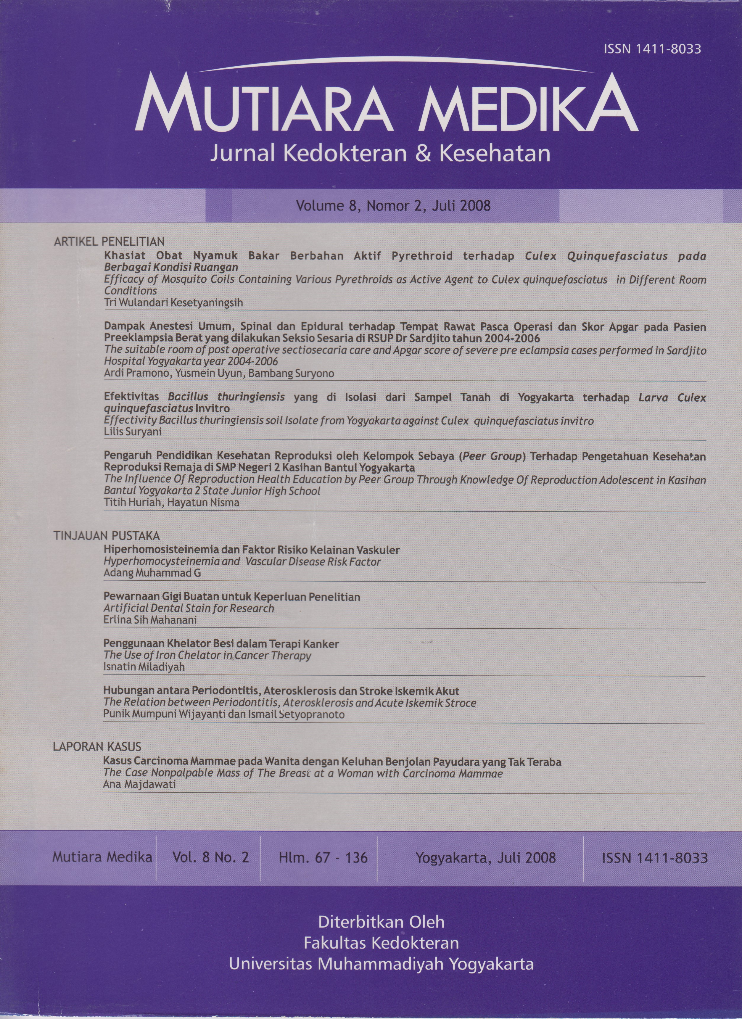
Kasus Carcinoma Mammae pada Wanita dengan Keluhan Benjolan Payudara yang Tak Teraba (Nonpalpable Mass) : Peran Ultrasonografi dan Mammografi sebagai Screening Diagnostik
DOI:
https://doi.org/10.18196/mmjkk.v8i2.1478Keywords:
Carcinoma ductal in situ, Carcinoma mammae, mammography, rise faktor, ultrasonography, Faktor risiko, Mammografi, Tumor ganas payudara, UltrasonografiAbstract
Breast neoplasma is the malignancy of the women that first number and caused highest mortality. We must think screening diagnostic to nonpalpable mass on the breast with have symptoms and rise facors. The case was diagnosed by anamnesis of the rise faktor, breast clinical examination, laboratory of BRCA1-2, imaging radiology and histopathology examination. Reported a woman, 44 years old, was menarche on 11 years old, pain in the left breast until upper extremity since two month ago. The craniocaudal and mediolateral position mammography were found multiple linier microcalsification in medioinferior aspect of left breast and not seen low/high density lession around it. Ultrasonography examination was found hypo echoic solid lesion, irreguler shape, ill define, weidht compare deep upper 1. The result of combination mammography and ultrasonography is suspect malignancy mass (BIRADS IV). Patient was operated and sent biopsy of the breast to examine histopathologic Anatomy laboratory. The result histopatholy is find Carcinoma ductal in situ and continued Modified Radical Mastectomy operation at all left breast. Then, the patient is continued chemoterapy and radioterapy treatment. The conclusion of this case report point that imaging radiology is important to diagnose screening. The mammography apperance is microcalsification and the ultrasound seem the hypoechoic lesion with ill define and deep per weidht one more pointed breast malignancy tumour. The Validity combination mammography and ultrasound high enough, with 91% of sensitivity and 98% of specivicity.. The early finding of breast screening can increase five survival rate in the patient.
Tumor ganas payudara merupakan keganasan pada wanita yang menduduki peringkat teratas dan sebagai penyebab kematian yang tinggi. Tumor ganas payudara dini kadang tidak memberikan gej ala berupa terabanya massa (nonpalpable mass), sehingga perlu dipikirkan screening diagnostik dengan mempertimbangkan berbagai faktor risiko dan gejala klinis yang mendukung. Penegakan diagnosis pada kasus keganasan pada payudara meliputi anamnesis dengan menggali faktor risiko, pemeriksaan fisik payudara, laboratorium (BRCA.j 2), pemeriksaan penunjang radiologi dan histopatologi. Dilaporkan wanita, 44 tahun dengan menarche 11 tahun, keluhan payudara kiri nyeri dijalarkan sampai lengan dan puting lecet selama 2 bulan. Hasil pemeriksaan mammografi posisi craniocaudal (CC) danmediolateral oblique (MLO) didapatkan mikrokalsifikasi linier, 2 buah di aspekmedioinferior dan tak tampak lesi densitas tinggi/rendah pada kedua payudara. Hasil pemeriksaan ultrasonografi, tampak lesi solid hypoechoic dengan bentuk irreguler, batas tak tegas irreguler, perbandingan tebal dan lebar lesi lebih dari 1. Hasil kombinasi pemeriksaan ultrasonografi dan mammografi mengarahkan lesi malignancy sesuai BIRADS IV. Penderita dilakukan operasi dan durante operasi dilakukan biopsi jaringan yang dilanjutkan pemeriksaan histopatologi. Hasil pemeriksaan histopatologi menunjukkan carsinoma ductal insitu dan pada puting yang lecet juga menunjukkan sel ganas, sehingga dilakukan pengangkatan payudara kiri seluruhnya (Modified Radical Mastectomy) dilanjutkan terapi radiasi dan chemoterapi. Kasus ini menujukkan bahwa peran pencitraan radiologi untuk screening diagnosis kelainan payudara sangat penting. Gambaran mikrokalsifikasi pada mammografi dan lesi hypoechoic, batas tak tegas dengan perbandingan deep/weidht lebih dari 1 mengarahkan pada suatu keganasan dengan validitas diagnostik tinggi (sensitifitas 91% dan spesifisitas 98%). Deteksi dini terutama pada kasus nonpalpable mass pada payudara akan memperbaiki keberhasilan terapi dan meningkatkan angka harapan hidup penderita.
References
Anonim, 2004, Data Tabulasi Dasar Rawat Inap Seluruh Rumah Sakit di Indonesia, Pelayanan Medis, Departemen Kesehatan RI.
Skinner, K.A, et al, 2001. Palpable Breast Cancers Are Inherently Different From Nonpalpable Breast Cancers, Annals of Surgical Oncology, 8(9):705- 710.
Thomas, A.G., Jermal, A. & Thun, M.J., 2004. Breast Cancer, Fact and Figure 2001-2002, the American Cancer Society, Atlanta, Georgia.
Forsberg, F., Merrit.C.R.B., Parker,L, Maitino.A.J., Merton, D.A., et al, 2004. Diagnosing Breast Lesions with Contrast-Enhance 3-Dimensional Power Doppler Imaging. J.Ultrasound Med. 23:173-182.
Houssami, N., Irwig, W., Simpson.J.M/ ., McKessar, M., Blome, S.,Noakes, J., 2003 Sydney Breast Imaging Accuracy Study: Comparative Sensitivity and Specificity of Mammografi and Sonography in Young Women with Symptoms, A JR ; 180:935-40.
Agustiningsih, S.R., 2004. Imaging Strategy in Breast Cancer Diagnostic, dalam Indonesian Issues on Breast Cancer, Surabaya.
Suprabawati, D.G.A., 2004. Clinical Examination and the Importance of Team Work Diagnosing (early) Breast Cancer, dalam Indonesian Issues on Breast Cancer 1, Surabaya.
Tjindarbumi, D., 2004. Penanganan Kanker Payudara Masa Kini dengan Berbagai Macam Issue di Indonesia, dalam Indonesian Issues on Breast Cancer 1, Surabaya,
Dershaw, D. 1997. Patient Selection and Management with Core Breast Biopsy. American Pathologist Breast J. 47:171-190
Shetty.M.K., Shah, Y.P. & Sharman, R.S., 2003. Prospective Evaluation of the Value of Combined Mammographic and Sonographic Assesment in Patients with Palpable Abnormalities of the Breast, J Ultrasound Med 22:263-268.
Soo, M.S., Rosen,E.L., Baker, J.A., Vo ,T.T. & Boyd ,B.A., 2001. Negative Predictive Value of Sonography with Mammografi in patients with Palpable Breast Lesions, AJR: 177, November.
Fewig, S.A., Picolli, C.W. 1997. The Breast in : Grainger & Allison’s Diagnostic Radiology, A.Textbook of Medical Imaging. Churchill Livingstone . New York . page 1995-2021
Parikh, J.R., Porter,B. Understanding Breast Ultrasound. 2002. www.imaainaeconomics.com.htm
Singhal, H., Thomson, S. 2004. Breast Cancer Evaluation. www.eMedicine.com
Anonim. 2005. Nuclear Medicine Breast Imaging (Scintimammografi).
Dongola, N., 2005. Breast Cancer Mammografi. Departement of Radiology, Sobo University Hospital.
Gunderman, R.B., 2006, Essenstial Radiology, 2nd ed, Thieme, New York.
Meschan, I. & Bertrand, M.L., 1987. Radiology of the Breast, in Meschan, I., Roentgen Signs in Diagnostic Imaging, Volume 4, W.B. Saunders Company.
Yang.W.T. & Tse.G.M.K. 2004. Sonographic, Mammographic and Histopathologic Corellation of Symptomatic Ductal Carcinoma In Situ, AJR; 182:101-110.
Yang, W.T., Suen, M., Ahuja, A.,
Metrewli, C., 1997, In Vivo Demontration of Microcalcification in Breast Cancer Using High Resolution Ultrasound, BrJ Rad. 70: 685-90
Tabar, L., Dean, B.P. 1985, Analysis of Calcification in : Teaching Atlas of Mammografi. Ed 2. Stuttgart New York. 137-170
Zonderland, H.M., Coerkamp, E.G., Herman, J., Vijver, M.J. & Voorthuisen, A.E., 1999. Diagnosis of Breast Cancer: Contribution of US as an Adjunct to Mammografi, Radiology, 213:413-422.
Downloads
Published
Issue
Section
License
Copyright
Authors retain copyright and grant Mutiara Medika: Jurnal Kedokteran dan Kesehatan (MMJKK) the right of first publication with the work simultaneously licensed under an Attribution 4.0 International (CC BY 4.0) that allows others to remix, adapt and build upon the work with an acknowledgment of the work's authorship and of the initial publication in Mutiara Medika: Jurnal Kedokteran dan Kesehatan (MMJKK).
Authors are permitted to copy and redistribute the journal's published version of the work (e.g., post it to an institutional repository or publish it in a book), with an acknowledgment of its initial publication in Mutiara Medika: Jurnal Kedokteran dan Kesehatan (MMJKK).
License
Articles published in the Mutiara Medika: Jurnal Kedokteran dan Kesehatan (MMJKK) are licensed under an Attribution 4.0 International (CC BY 4.0) license. You are free to:
- Share — copy and redistribute the material in any medium or format.
- Adapt — remix, transform, and build upon the material for any purpose, even commercially.
This license is acceptable for Free Cultural Works. The licensor cannot revoke these freedoms as long as you follow the license terms. Under the following terms:
Attribution — You must give appropriate credit, provide a link to the license, and indicate if changes were made. You may do so in any reasonable manner, but not in any way that suggests the licensor endorses you or your use.
- No additional restrictions — You may not apply legal terms or technological measures that legally restrict others from doing anything the license permits.


