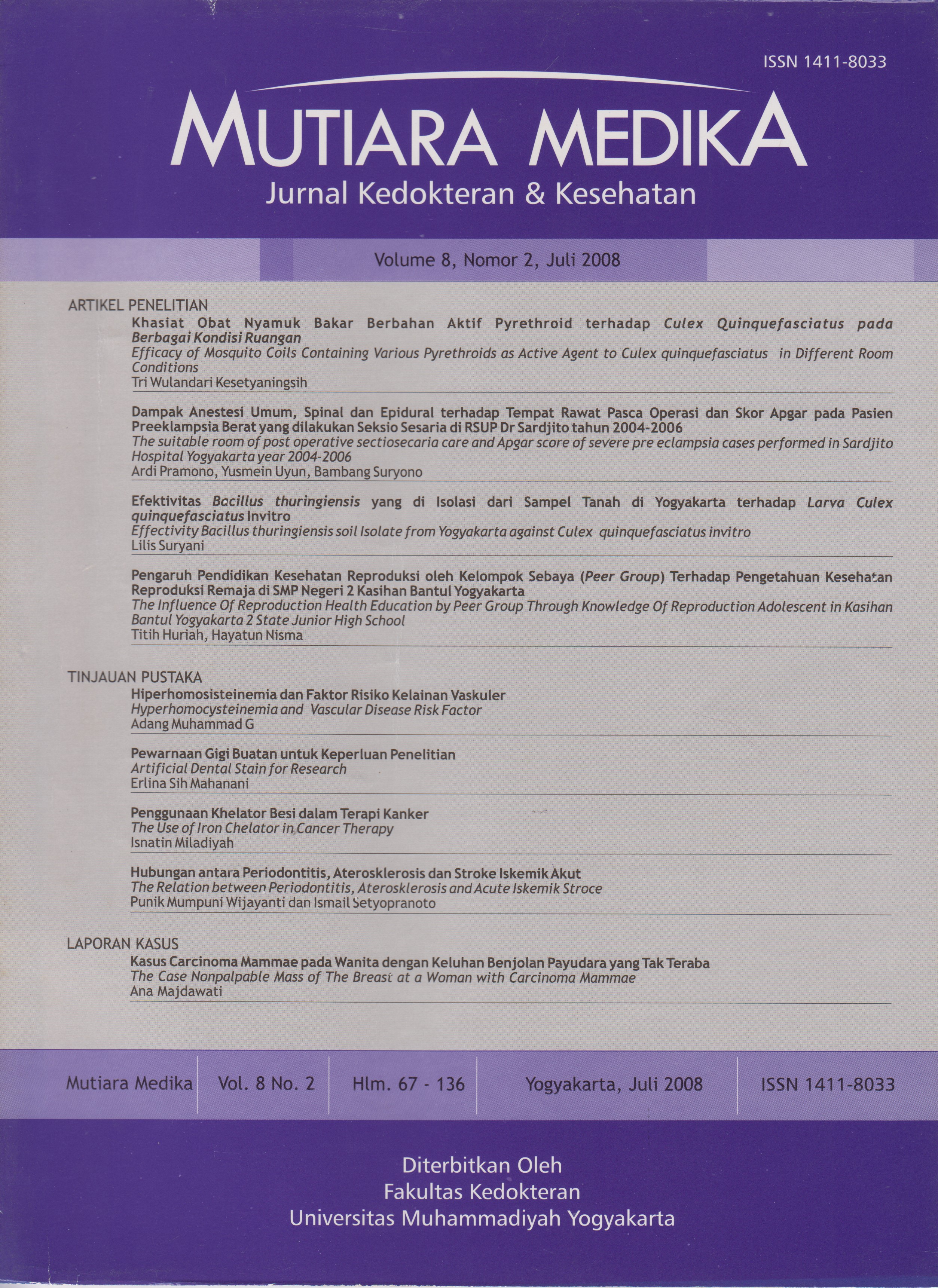
Penggunaan Khelator Besi dalam Terapi
DOI:
https://doi.org/10.18196/mmjkk.v8i2.1484Keywords:
carcinogenesis, iron, iron chelator, besi, karsinogenesis, khelator besi, terapi kankerAbstract
Iron is essential for numerous crucial biochemical reactions ranging from cellular respiration in mitochondria to DNA synthesis. Neoplastic cells have a high iron requirement because of their rapid rate ofproliferation. The close linkage between cell proliferation and iron lead to suggestion that iron deprivation could be a useful strategy for inhibition of tumor cell growth. In vitro and in vivo studies showed that iron chelator desferrioxamine (DFO) that has been traditionally used in the treatment of iron overload, showed ability to limit growth of tumor cells. Iron chelators demonstrated ability to inhibit tumor cell growth by their activity to induce apoptosis and inhibit cell cycle progression, particularly the G/S transition. This article reviews role of iron in carcinogenesis and mechanism of actions of iron chelators in the treatment of cancer.
Besi berperan dalam berbagai reaksi biokimia penting mulai dari respirasi seluler di mitokondria sampai sintesis DNA. Sel-sel neoplasma membutuhkan besi dalam jumlah besar karena pertumbuhannya cepat. Hubungan erat antara proliferasi sel dengan besi menimbulkan pemikiran bahwa pengurangan kadar besi mungkin dapat menjadi salah satu strategi dalam menghambat pertumbuhan tumor. Khelator besi desferioksamin (DFO) yang secara tradisional telah digunakan secara luas untuk terapi keracunan besi ternyata mampu menghambat pertumbuhan berbagai sel kanker baik in vitro maupun in vivo. Khelator besi mampu menghambat pertumbuhan tumor melalui induksi apoptosis dan hambatan pada siklus sel terutama pada fase G/S. Tulisan ini bertujuan untuk menelaah lebih lengkap mengenai peran besi dalam proses karsinogenesis dan bagaimana mekanisme keija khelator besi dalam terapi kanker.
References
Yoon G., Kim H.J., Yoon Y.S., Cho H., Lim I.K, and Lee J.H. 2002. Iron cheiation-induced senescence-like growth arrest in hepatocyte cell lines: association of transforming growth factor |31 (TGF-j31)-mediated p27Kip1 expression. Biochem J 366: 613-621
Andreau G.P., Delgado R., Velho J.A., Curti C., and Vercesi AE. 2005. Iron complexing activity of mangiferin, a naturally occuring glucosylxanthine, inhibits mitochondrial lipid peroxidation induced by Fe2+-citrate. EurJofPharm 513: 47-55
Huang X. 2003. Iron overload and its association with cancer risk in humans: evidence for iron as a carcinogenic metal. Mutation Res 533: 153-171
Abeysinghe R.D., Greene B.T., Haynes R., Willingham M.C., Turner J., Planalp R.P., Brechbiel M.W., Torti F.M., and Torti S.V. 2001. p53-independent apoptosis mediated by tachpyridine, an anticancer iron chelator. Carcinogenesis 22 (10): 1607-1614
Zhao Y dan Xu JX. 2004. The operation of alternative electron-leak pathways mediated by cytochrome c in mitochondria. Biochem and Biophys Res Comm 317: 980-987
Hann HMW. 2002. Prevention on Hepatocellular Carcinoma: antiviral therapy, preneoplastic markers and iron nutrition. The Korean Journal of Gastroenterology 39:105-109
Nyholm, S., Mann, G.J., Johansson, A.G., Bergeron, R.J., Graslund, A., and Thelander, L. 1993. Role of Ribonucleotide Reductase in Inhibition of Mammalian Cell Growth by Potent Iron Chelators. J of Biol Chem 268 (35): 26200-26205
Chaston T.B., Lovejoy, D.B., Watts, R.N., and Richardson D.R. 2003. Examination of the Antiproliferative Activity of Iron Chelators: Multiple Cellular Targets and the Different Mechanisms of Action of Triapine Compared with Desferrioxamine and the Potent Pyridoxal Isonicotinoyl Hydrazone Analogue 311. Clin Cancer Res 9: 402-414
Bouzyk E., Gradzka I., Iwanenko T, Kruszewski M., Sochanowicz B., and Szumiel I. 2000. The response of L5178Y lymphoma sublines to oxidative stress: Antioxidant defence, iron content and nuclear translocation of the p65 subunit of NF-KB. Acta Biochimica Polonica 47 (4): 881-888
Cragg, L, Hebbel, R.P., Miller, W., Solovey, A., Selby, S., and Enright, H. 1998. The iron Chelator L1 Potentiates Oxidative DNA Damage in Iron-Loaded Liver Cells. Blood 92 (2): 632-638
Xiong S., She H., Takeuchi H., Han B., Engelhardt J.F., Barton C.H., Zandi E., Giulivi C., and Tsukamoto H. 2003. Signaling Role of Intracellular Iron in NF- KB Activation. J of Biol Chem 278 (20): 17646-17654
Sellers, W.R, and Fisher, D.E. 1999. Apoptosis and cancer drug targeting. J of Clin Invest 104 (12): 1655-1661.
Harris G.K, and Shi X. 2003. Signaling by carcinogenic metals and metal- induced reactive oxygen species. Mutation Res 533: 183-200
Simonart, T. 2004. Iron: a target for the management of Kaposi’s sarcoma? BMC Cancer 4 (1)
Gao J. and Richardson D.R. 2001. The potential of iron chelators of the pyridoxal isonicotinoyl hydrazone class as effective antiproliferative agents, IV: the mechanisms involved in inhibiting cell-cycle progression. Neoplasia 98 (3): 842-850
Parton, M., Dowsett, M., and Smith, I. 2001. Studies of apoptosis in breast cancer. BMJ 322: 1528-1532
Collins K., Jacks T, and Pavletich N.P. 1997. The cell cycle and cancer. Proc Natl Acad Sci USA 94: 2776-2778
Rakba, N., Loyer, R, Gilot, D., Delcros, J.G., Glaise, D., Baret, R, Pierre, J.L., Brissot, R, and Lescoat, G. 2000. Antiproliferative and apoptotic effects of O-Trensox, a new synthetic iron chelator, on differentiated human hepatoma cell lines. Carcinogenesis 21 (5): 943-951
Narla R.K., Chen C.L., Dong Y, and Uckun F.M. 2001. In vivo Antitumor Activity of Bis(4,7-dimethyl-1,10- phenantroline) Sulfatooxovanadium (IV) {METVAN [V0(S04)(Me2-Phen)2]}. Clin Cancer Res 7: 2124-2133
Sam M., Hwang J.H., Chanfreau G, and Abu-OmarM.M. 2004. Hydroxyl Radical is the Active Species in Photochemical DNA Strand Scission by Bis(peroxo)vanadium (V) Phenanthroline. Inorg Chem 43: 8447- 8455
KicicA., ChuaACG., and Baker E. 2001. Effect of Iron Chelators on Proliferation and Iron Uptake in Hepatoma Cell. Cancer 92(12): 3093-3110
KicicA., and Baker E. 1998. The Effects of Iron Chelator Upon Cellular Proliferation and Iron Transport in Hepatoma Cells. Presented at I NAB IS ’98 - 5th Internet World Congress on Biomedical Sciences at McMaster University, Canada, Dec 7-16th. Invited Symposium.
Greene, B.T., Thorburn, J., Willingham, M.C., Thorburn, A., Planalp, R.P., Brechbiel, M.W., Jennings-Gee, J., Wilkinson IV, J., Torti, F.M., and Torti, S.V. 2002. Activation of Caspase Pathway during Iron Chelator-mediated Apoptosis. J of Biol Chem 277 (28): 25568-25575
Torti S.V., Torti F.M., Whitman S.P., Brechbiel M.W., and Planalp R.P. 1998. Tumor Cell Cytotoxicity of a Novel Metal Chelator. Blood 92 (4): 1384-1389
Yuan J., Lovejoy D.B., and Richardson D.R. 2004. Novel di-2-pyridyl-derived iron chelators with marked and selective antitumor activity: in vitro and in vivo assessment. Blood 104 (5): 1450-1458
Alcain F.J., Low H., and Crane F.L. 1994. Iron reverses impermeable chelator inhibition of DNA synthesis in CCI 39 cells. Proc Natl Acad Sci USA 91:7903-7906
Planalp RR, Przyborowska AM., Park G., Ye N., Lu FH., Rogers RD., Broker GA., Torti SV, and Brechbiel MW. 2002. Novel cytotoxic chelators that bind iron(ll) selectively over zinc(ll) under aqueous aerobic conditions. Biochem Soc Transactions 30 (4): 758-762
Downloads
Published
Issue
Section
License
Copyright
Authors retain copyright and grant Mutiara Medika: Jurnal Kedokteran dan Kesehatan (MMJKK) the right of first publication with the work simultaneously licensed under an Attribution 4.0 International (CC BY 4.0) that allows others to remix, adapt and build upon the work with an acknowledgment of the work's authorship and of the initial publication in Mutiara Medika: Jurnal Kedokteran dan Kesehatan (MMJKK).
Authors are permitted to copy and redistribute the journal's published version of the work (e.g., post it to an institutional repository or publish it in a book), with an acknowledgment of its initial publication in Mutiara Medika: Jurnal Kedokteran dan Kesehatan (MMJKK).
License
Articles published in the Mutiara Medika: Jurnal Kedokteran dan Kesehatan (MMJKK) are licensed under an Attribution 4.0 International (CC BY 4.0) license. You are free to:
- Share — copy and redistribute the material in any medium or format.
- Adapt — remix, transform, and build upon the material for any purpose, even commercially.
This license is acceptable for Free Cultural Works. The licensor cannot revoke these freedoms as long as you follow the license terms. Under the following terms:
Attribution — You must give appropriate credit, provide a link to the license, and indicate if changes were made. You may do so in any reasonable manner, but not in any way that suggests the licensor endorses you or your use.
- No additional restrictions — You may not apply legal terms or technological measures that legally restrict others from doing anything the license permits.


