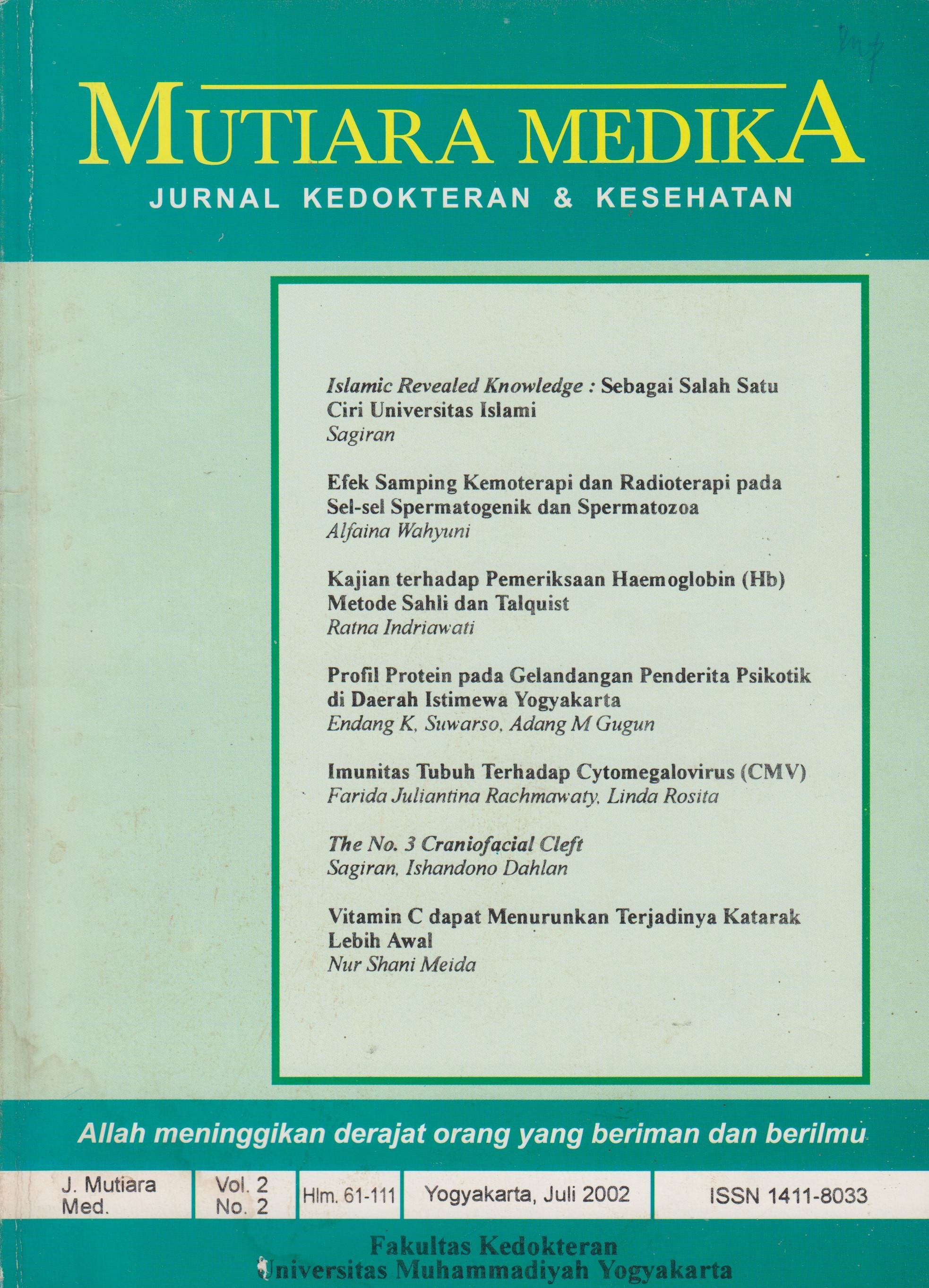
Efek Samping Kemoterapi dan Radioterapi pada Sel-sel Spermatogenik dan Spermatozoa
DOI:
https://doi.org/10.18196/mmjkk.v2i2.1507Keywords:
radiotherapy, chemotherapy, spermatogonia, radioterapi, kemoterapiAbstract
Medical treatment for cancer is a combination of operative treatment, ra-diotherapy and chemotherapy. Theoretically, anticancer agent can kill can¬cer cells. However, it also causes many side effects especially on the normal cells, which have high mitosis activity. One of them is spermatogenic cell. Anticancer agent is included into reproductive toxin. Its working mecha-nism is by the alkylation of biologic molecules. Radiotherapy and chemotherapy reduce the number of spermatogonia Al, spermatogonia B and cause aberation of DNA structures on the next-generation cells including spermatozoa, hence result in the decrease of number of spermatozoa and sperm motility and the increase of the percentage of spermatozoa with abnormal morphology. The effects of radiotherapy and chemotherapy are temporary and reversibel. The cell recovery depends on the type of anticancer agent, its dosage and the length of therapy applied. Spermatogonia stem cells are the most important factors on this process.
Tindakan medis yang dilakukan untuk pengobatan kanker adalah kombinasi pembedahan, radioterapi dan kemoterapi. Secara teoritis bahan antikanker bisa membunuh sel kanker, tetapi kenyataannya banyak menimbulkan efek samping terutama pada sel normal yang mempunyai aktivitas pembelahan cepat. Salah satu diantaranya adalah sel-sel spermatogenik. Bahan-bahan antikanker termasuk dalam golongan toksin reproduktif. Mekanisne kerjanya dengan cara mengalkilasi molekul biologis. Pascaradioterapi dan kemoterapi terjadi penurunan jumlah spermatogonia Al dan B dan menyebabkan aberasi struktur DNA pada generasi sel berikutnya termasuk spermatozoa. Akibatnya jumlah dan motilitas menurun dan persentase abnormalitas spermatozoa meningkat. Efek radioterapi dan kemoterapi bersifat temporer dan bisa terjadi pemulihan. Pemulihan sangat tergantung pada jenis bahan antikanker, dosis dan lama pemberian. Sel spermatogonia induk merupakan faktor terpenting dalam proses tersebut.
References
Amelar,R.D.,Dubin,L., and Walsh,P.C. 1977. Male Infertiliy. W.B. Saunders Company. Philadelphia.
Bardin,C.W.,Cheng,C.Y.,Musto,N.E., and Gunsalus,G.L. 1988. The Sertoli Cell, in: Knobil,E, and Neil,J. (eds). The Phisiology of Reproduction. Raven Press,Ltd., New York.
Bedford, J.M. 1975. Maturation, Transport and Fate of Spermatozoa in the Epididymis, in: Greep, R.O. Astwood E.B., Hamilton, D.W. and Geiger, S.R. (eds). Handbook of Physiology, Section Endocrinology vol. V. Waverly Press, Inc., Baltimore.
Chaterjee,R., Haines,G.A., Perers,D.M., Goldstone,A. and Morris, I.D. 2000. Testicular and Sperm DNA Damage after Treatment with Fludarabine for Chronic Lymphocytic Leukaemia. Hum- Reprod. 15(4): 762-7.
du Pan,M.R. and Campana. 1993. Phisiopathology of Spermatogenic Arrest. Fertil. Steril. 60 (6):937-51
Hadley, M.E. 1992. Endocrinology. Prentice Hall International, London.
Heyzer. 1987. Obat Antikanker. Dalam Farmakologi dan Terapi. FKUI. Jakarta
Howell,S. and Shalet,S. 1998. Gonadal Damage from Chemotherapy and Radiotherapy. Endocrinol. Metab. Clin. North. Am. 27(4): 927 - 43.
Meistrich, M.L. 1986. Critical Components of Testicular Function and Sensitivity to Disruption. Biol. Reprod{34): 17-28.
Orgebin-Crist, M.C., Danzo, B.J. and Davies, J. 1975. Endocrine Control of The Development and Maintenance of Sperm Fertilizing Ability in The Epididymis, inm: Greep, R.O. Astwood E.B., Hamilton, D.W. and Geiger, S.R. (eds). Handbook of Physiology, Section Endocrinology vol. V. Waverly Press, Inc., Baltimore.
Downloads
Published
Issue
Section
License
Copyright
Authors retain copyright and grant Mutiara Medika: Jurnal Kedokteran dan Kesehatan (MMJKK) the right of first publication with the work simultaneously licensed under an Attribution 4.0 International (CC BY 4.0) that allows others to remix, adapt and build upon the work with an acknowledgment of the work's authorship and of the initial publication in Mutiara Medika: Jurnal Kedokteran dan Kesehatan (MMJKK).
Authors are permitted to copy and redistribute the journal's published version of the work (e.g., post it to an institutional repository or publish it in a book), with an acknowledgment of its initial publication in Mutiara Medika: Jurnal Kedokteran dan Kesehatan (MMJKK).
License
Articles published in the Mutiara Medika: Jurnal Kedokteran dan Kesehatan (MMJKK) are licensed under an Attribution 4.0 International (CC BY 4.0) license. You are free to:
- Share — copy and redistribute the material in any medium or format.
- Adapt — remix, transform, and build upon the material for any purpose, even commercially.
This license is acceptable for Free Cultural Works. The licensor cannot revoke these freedoms as long as you follow the license terms. Under the following terms:
Attribution — You must give appropriate credit, provide a link to the license, and indicate if changes were made. You may do so in any reasonable manner, but not in any way that suggests the licensor endorses you or your use.
- No additional restrictions — You may not apply legal terms or technological measures that legally restrict others from doing anything the license permits.


