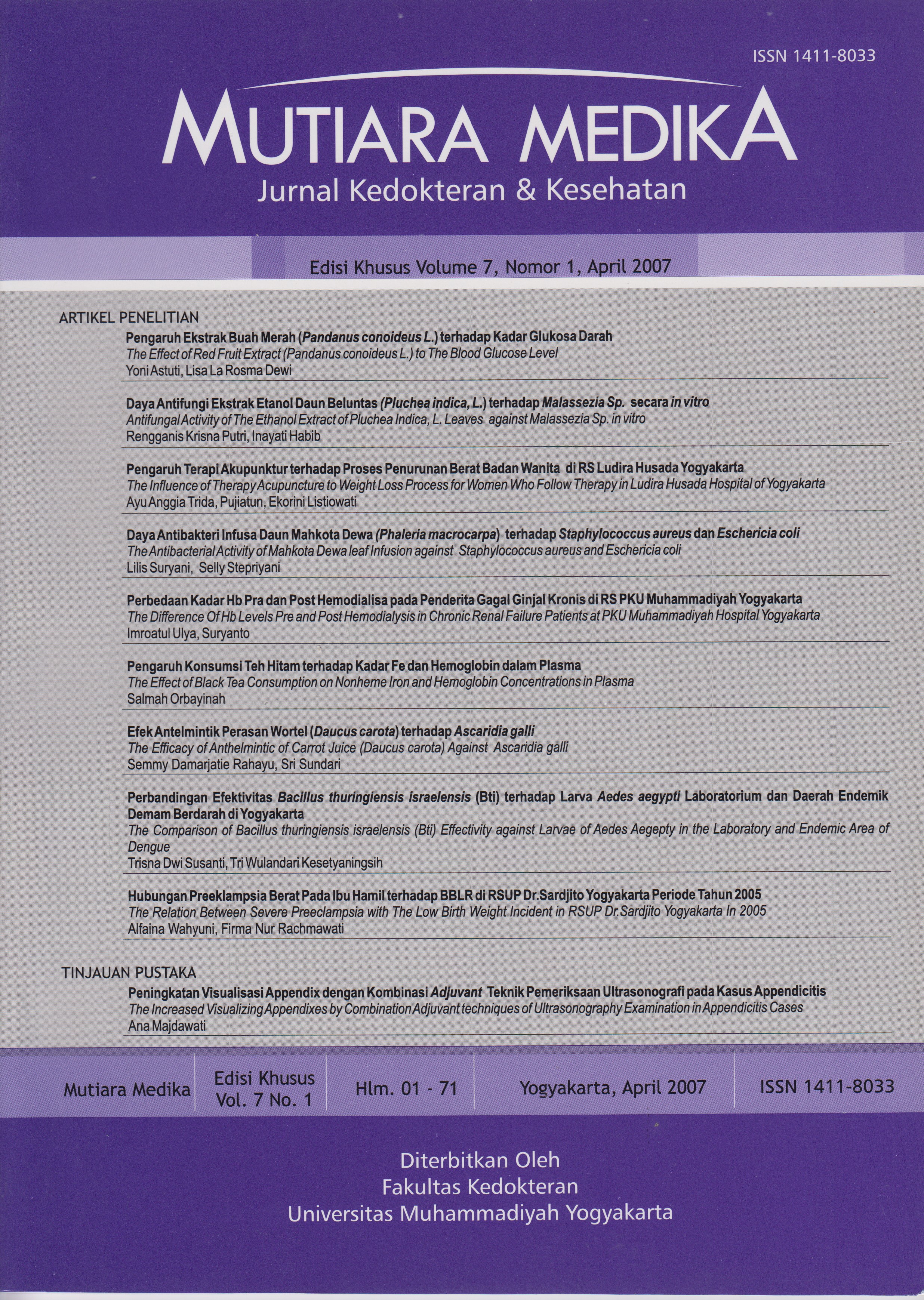
Peningkatan Visualisasi Appendix dengan Kombinasi Adjuvant Teknik Pemeriksaan Ultrasonografi pada Kasus Appendicitis
DOI:
https://doi.org/10.18196/mmjkk.v7i1%20(s).1686Keywords:
appendicitis, teknik graded kompressi, teknik adjuvant, akurasi diagnostik, diagnosis, adjuvant, USGAbstract
To diagnose the appendicitis not easy, because in the fact we find many false positive appendicitis post surgery. Ultrasonography is modality that often choose because it has severe superiority, i.e : non invasif, safety, no radiation effect (safety for pregnant woman and children), cheap, easy, simple and need short time. The weakness Ultrasonography is depend on the operator, various appendix location each somebody. The aim of this literature reviews is to resume of various operator-dependent techniques to graded compression ultrasonography that is useful for allowing improved visualization of appendixes. The addition of various operator-dependent techniques to graded compression sonography i.e a posterior manual compression technique, upward graded compression technique, left oblique lateral decubitus position of body, low-frequency convex transducer (especially for obese poeple and gravid woman), the combination of gray scale and color doppler ultrasound can increase diagnostic accuracy of appendicitis (sensitivity, specitivity and accuracy more 99%). The addition of adjuvant technique of dependent operator that have most highly accuracy is a posterior manual compression technique and the lowest is Left oblique lateral decubitus position of body. The result of the research find appendicitis appearances in ultrasound that have most highly accuracy are blind ending tubular structure at the transversal or longitudinal section of ultrasound, diameter of wallthichness more 6 mm, non compressible, non peristaltic, vascularity increased around appendixes in color doppler ultrasound examination.
Penegakan diagnosis appendicitis merupakan hal yang tidak mudah, karena kenyataannya di lapangan masih banyak angka positif palsu appendicitis post operasi. Ultrasonografi (USG) merupakan pemeriksaan yang paling sering dipilih sebagai modalitas diagnostik appendicitis karena mempunyai beberapa keunggulan, yaitu: non invasif, aman, efek radiasi tidak ada (aman bagi wanita hamil dan anak-anak), relatif murah dan mudah dilakukan, waktu yang diperlukan singkat. Kekurangannya pemeriksaan USG sangat tergantung ketrampilan pemeriksa, lokasi appendix yang bervariasi untuk tiap orang sehingga teknik pemeriksaan U SG sangat penting diperhatikan. Tujuan literature review ini adalah untuk merangkum berbagai teknik pemeriksaan USG untuk memvisualisasikan appendix sehingga dapat meningkatkan nilai diagnostik appendicitis. Berdasar literatur review ini dapat disimpulkan bahwa teknik graded kompresi dengan Adjuvant teknik pemeriksaan USG dependent operator yaitu: teknik manual posterior, teknik kompresi bertahap ke arah atas, teknik perubahan posisi Left Oblique Lateral Decubitus, penggunaan transducer konveks frekuensi rendah (terutama untuk orang yang obese dan wanita hamil), kombinasi USG gray scale dengan CDU dapat meningkatkan nilai akurasi diagnostik appendicitis (sensitifitas, spesifisitas dan akurasi hampir 99%). Beberapa teknik Adjuvant tersebut yang mempunyai nilai diagnostik tertinggi adalah teknik kompresi manual posterior, sedang yang terendah adalah teknik perubahan posisi tubuh (Left Oblique Lateral Decubitus). Gambaran USG pada appendix yang mengarah appendicitis yang mempunyai akurasi tinggi berdasar beberapa penelitian, adalah: tampak struktur tubular buntu (blind ending) pada potongan longitudinal dan transversal, diameter dinding lebih 6 mm, non compressible, aperistaltik, peningkatan aliran darah pada pemeriksaan CDU di daerah appendix.
References
Anonim; Appendicitis: Akute Abdomen and Surgical Gastroenterology
Craig, S, MD, 2006; Appendicitis Akute, www.Emedicine
Jacobs, J.E; 2006; CT and Sonography for Suspected Akute Appendicitis: A Commentary; AJR 2006: 186: 1094-1096
Kessler, N; Cysteval, C; Gallix, B; Lesnik, A; Blayac, P.M, et al; 2003, Appendicitis: Evaluation of Sensitivity, Specificity, and Predictive Values of US, Doppler US, and Laboratory Findings; Radiology 2004; 230: 472 - 478
Baldisserotto, M dan Marchiori, E; 2000; Accuracy of Noncompressive Sonography of Children with Appendicitis According to the Potential Positions of the Appendix; AJR 2000; 175: 1387-1392
Hwa Lee, J; Ki Joeng, Y; Bo Park, J, Kang Park, J; Jeong, A.K, et al; 2005; Operator Dependent Technique for Graded Compression Sonography to Detect the Appendix and Diagnose Akute Appendicitis; AJR 2005; 184: 91¬97
Hwa Lee, J; Ki Jeong, Y; Hwang ,J.C; Ham, S.Y; Yang, S.O; 2002; Graded Compression Sonography with Adjuvant Use of a posterior Manual Compression Technique in the Sonographic Diagnosis of Akute Appendicitis; AJR 2002; 178: 863-868
Rettenbacher, T; Hollerweger, A; Macheiner, P; Rettenbacher, L; Frass, R, et al; 2000; Presence or Absence of Gas in the Appendix: Additional Criteria to Rule Out or Confirm Akute Appendicitis Evaluation with US; Radiology 2000; 214: 183-187
Anonim; Diagnostic Ultrasound; http://www.medscape.com
Anonim; Coloproctology: Appendicitis
Santacroce, L; 2006; Appendicitis www.Emedicine
Anonim; Vermiform appendix; Wikipedia, the free encyclopedia.htm.
Puylaert, J. B.C.M; 1986; Acute Appendicitis: US Evaluation Using Graded Compression ; Radiology 1986; 158:355-360.
Bergman, R.A, et al , Atlas of Microscopic Anatomy: Section 10 - Digestive System www.anatomvatlases.org
Lim, K.H; J.Lee, W; J.Lee, S; Namgung, S; Lim, J.H; 1996; Focal Appendicitis Confined to the Tip Diagnosis at US; Radiology 1996; 200: 799-801
Chesbrough, R.M; Burkhard, T.K; Balsara, Z.N; Goff, W.B; Davis, D.J; 1993; Self Localization in US of Appendicitis: An Addition to Graded Compression; Radiology 1993; 187: 349-351
Quillin,S.P; Siegel, M.J; 1994, Appendicitis: Efficacy of Color Doppler Sonography; Radiology 1994 ; 191 : 557-560
Anonim; Diagnostic Imaging: Ultrasound: Technique and Accuracy; file://H:AnatomiAppendicitis.htm
Craig, S, 2006, Appendicitis Acute, in http:// www. eMedicine
Rettenbacher, T; Hollerweger, A; Macheiner, P; Gritzmann, N; Daniaux, M, et al, 2003; Ovoid Shape of the Vermiform Appendix: A Criterion to Exclude Akute Appendicitis Evaluation with US; Radiology 2003; 226:95-100
Shawn P.Q, et al, 1994, Appendicitis acut, Radiology 1994 ; 191 : 557-560
Downloads
Published
Issue
Section
License
Copyright
Authors retain copyright and grant Mutiara Medika: Jurnal Kedokteran dan Kesehatan (MMJKK) the right of first publication with the work simultaneously licensed under an Attribution 4.0 International (CC BY 4.0) that allows others to remix, adapt and build upon the work with an acknowledgment of the work's authorship and of the initial publication in Mutiara Medika: Jurnal Kedokteran dan Kesehatan (MMJKK).
Authors are permitted to copy and redistribute the journal's published version of the work (e.g., post it to an institutional repository or publish it in a book), with an acknowledgment of its initial publication in Mutiara Medika: Jurnal Kedokteran dan Kesehatan (MMJKK).
License
Articles published in the Mutiara Medika: Jurnal Kedokteran dan Kesehatan (MMJKK) are licensed under an Attribution 4.0 International (CC BY 4.0) license. You are free to:
- Share — copy and redistribute the material in any medium or format.
- Adapt — remix, transform, and build upon the material for any purpose, even commercially.
This license is acceptable for Free Cultural Works. The licensor cannot revoke these freedoms as long as you follow the license terms. Under the following terms:
Attribution — You must give appropriate credit, provide a link to the license, and indicate if changes were made. You may do so in any reasonable manner, but not in any way that suggests the licensor endorses you or your use.
- No additional restrictions — You may not apply legal terms or technological measures that legally restrict others from doing anything the license permits.


