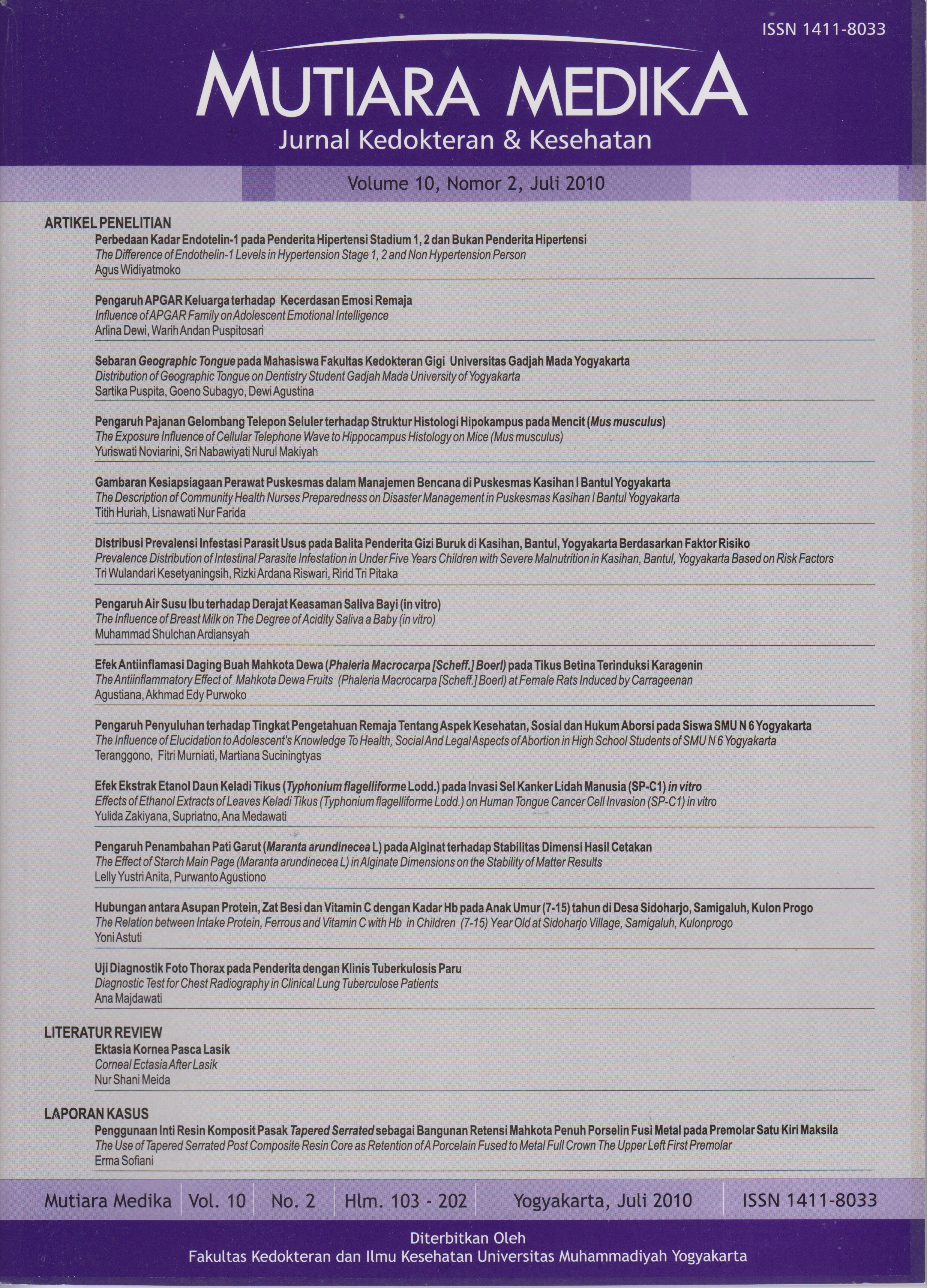
Uji Diagnostik Gambaran Lesi Foto Thorax pada Penderita dengan Klinis Tuberkulosis Paru
DOI:
https://doi.org/10.18196/mmjkk.v10i2.1582Keywords:
tuberkulosis paru, gejala klinik, foto thorax, uji diagnostik, lung tuberculose, clinical sign, chest radiography, diagnostic testAbstract
Chest radiography is one of examination to diagnose lung TB. More lesions in chest radiography as infiltrate, fibroinfiltrate, cavitas, calcification, pleural effusion, etc. often find in chronic lung diseases, especially lung TB. The aim of this study to determine the sensitivity and spesificity of clinical symptoms and chest x-ray lesions. This is a retrospective study of medical record of polyclinic and hospitalized patient of Bantul District Hospital year 2010. Diagnostic test research methods are based on the gold standard smear of sputum. There are 100 samples, consisting of 50 with clinical TB and 50 without, aged 18-50 year old with chest X-ray and sputum smear examination. The result showed the most clinical symptoms of TB are bloody cough and shortness of breath. Photo radiography obtained 33 patients with lesions infiltrates, 18 patients a combination of more than 3 lesions, 4 patients with fibroinfiltrate and 45 patients without lesions. Sensitivity and specificity of clinical symptoms of TB 74.5%, 75.5%, photo- fibroinfiltrate chest infiltrates 83.3%, 24.4% and a combination of more than 3 lesions 87.5%, 13.3%. Summing up the sensitivity of clinical symptoms, infiltrates-fibroinfiltrate and a combination of more than 3 lesions is quite high (> 70%), whereas low specificity (<70%).
Foto thorax merupakan salah satu penunjang diagnostik tuberkulosis (TB). Lesi pada foto thorax seperti infiltrat, fibrosis, kalsifikasi, kavitas, effusi pleura maupun kombinasi lesi sering dijumpai pada penyakit radang kronik paru, terutama TB. Tujuan penelitian ini untuk mengetahui sensitifitas dan spesifisitas gejala klinis dan lesi foto thorax. Penelitian ini bersifat retrospektif dari catatan medik poliklinik dan bangsal RSUD Bantul tahun 2010. Ada 100 sampel, terdiri 50 dengan klinis TB dan 50 tanpa klinis TB, usia 18-50 tahun dengan foto thorax dan pemeriksaan sputum BTA. Metode penelitian uji diagnostik ini didasarkan pada baku emas sputum BTA. Hasil menunjukkan gejala klinis TB terbanyak adalah batuk berdarah dan sesak napas. Foto thorax didapatkan 33 pasien dengan lesi infiltrat, 18 pasien kombinasi lebih dari 3 lesi, 4 pasien dengan fibroinfiltrat dan 45 pasien tanpa lesi. Sensitifitas dan spesifisitas gejala klinis TB 74,5%, 75,5%, foto thorax infiltrat-fibroinfiltrat 83,3%, 24,4% dan kombinasi lebih 3 lesi 87,5%, 13,3%. Disimpulkan sensitifitas gejala klinis, infiltrat-fibroinfiltrat dan kombinasi lebih dari 3 lesi cukup tinggi (> 70%), sedangkan spesifisitasnya rendah (< 70%).
References
RI Sekretariat Jenderal Kementerian Kesehatan PENGENDALIAN TB DI INDONESIA MENDEKATI TARGET MDG [Online] // http://www.depkes. go. id/. - Kementerian Kesehatan Republik Indonesia, 2010. - 16 Oktober 2010. - http://www.depkes.go.id/index.php/ berita/press-release/857-pengendalian- TB-di-indonesia-mendekati-target-mdg. html.
Kasus TBC Naik hingga 75 Persen [Online] // Kompas.com. - Kompas, 19 Maret 2010. - 16 Oktober 2010 http://kesehatan.kompas.com/ read/2010/03/19/13472241/Kasus. TBC.Naik.hingga 75.%
Arifin dan Nawas, 2009. Diagnosis dan Penatalaksanaan TB Paru. Jakarta : Divisi Infeksi, Departemen Pulmonologi dan Ilmu Kedokteran Respirasi FKUI/ SMF Paru
Schiffman George Tuberkulosis (TB) [Online] // MedicineNet.com. - WebMD, 14 Agustus 2007. - 17 10 2010. - http:// www.medicinenet.com/Tuberkulosis/ article.htm. International Standard of Tuberkulosis Care, 2007
Centers for Disease Control and Prevention. Prevention and treatment of Tuberkulosis among patients infected with human immunodeficiency virus: Principles of therapy and revised recommendations. MMWR 1998;47 (RR-20): 1-58.
Icksan., Aziza. G., Luhur dan Reny. 2008. Radiologi Thorax Tuberkulosis Paru. Jakarta: Sagung Seto.
Amin, Z., Bahar., A. 2007. Buku Ajar Ilmu Penyakit Dalam. Jakarta: FKUI.
Alsagaff, H., Mukty, A.. 2006. Dasar- dasar Ilmu Penyakit Paru. Surabaya: Airlangga University Press.
Lemeshow S., Hosmer, D and Lwanga S. 1990. Adequacy of Sample Size for Health Studies. John Wiley & Sons, Chichester
Julie.M., Marteen.B., Joseph.S., Paulin.B., et al, 2008. Accuracy of Clinical Signs in The Diagnosis of Pulmonary Tuberkulosis: Comparison of Three Reference Standards Closing Data from a Tertiary Care Centre in Rwanda, in The Open Tropical Medicine Journal, 2008, I, 1-7
Anonim, PedomanNasionalT uberkulosis Anak, ed 2, UKK Respirology PP Ikatan Dokter Anak Indonesia, 2008
Gunderman, R.B., 2006. Essential Radiology, Clinical Presentation- Pathophysiology-Imaging: Respiratory System, ed 2, Thieme, New York, Stuttgart, page 68
Chapman, S and Nakiely, R., 1988. Aids to Radiological Differential Diagnosis, Bailliare Tindall
Rosadi I. 2004. Uji Sensitifitas dan Spesifisitas Pemeriksaan Darah dan Rontgen Thorak untuk DiagnosisTuberkulosis Paru di RSUD dr Soesilo di Slawi, Kabubaten Tegal, 2004
Downloads
Published
Issue
Section
License
Copyright
Authors retain copyright and grant Mutiara Medika: Jurnal Kedokteran dan Kesehatan (MMJKK) the right of first publication with the work simultaneously licensed under an Attribution 4.0 International (CC BY 4.0) that allows others to remix, adapt and build upon the work with an acknowledgment of the work's authorship and of the initial publication in Mutiara Medika: Jurnal Kedokteran dan Kesehatan (MMJKK).
Authors are permitted to copy and redistribute the journal's published version of the work (e.g., post it to an institutional repository or publish it in a book), with an acknowledgment of its initial publication in Mutiara Medika: Jurnal Kedokteran dan Kesehatan (MMJKK).
License
Articles published in the Mutiara Medika: Jurnal Kedokteran dan Kesehatan (MMJKK) are licensed under an Attribution 4.0 International (CC BY 4.0) license. You are free to:
- Share — copy and redistribute the material in any medium or format.
- Adapt — remix, transform, and build upon the material for any purpose, even commercially.
This license is acceptable for Free Cultural Works. The licensor cannot revoke these freedoms as long as you follow the license terms. Under the following terms:
Attribution — You must give appropriate credit, provide a link to the license, and indicate if changes were made. You may do so in any reasonable manner, but not in any way that suggests the licensor endorses you or your use.
- No additional restrictions — You may not apply legal terms or technological measures that legally restrict others from doing anything the license permits.


