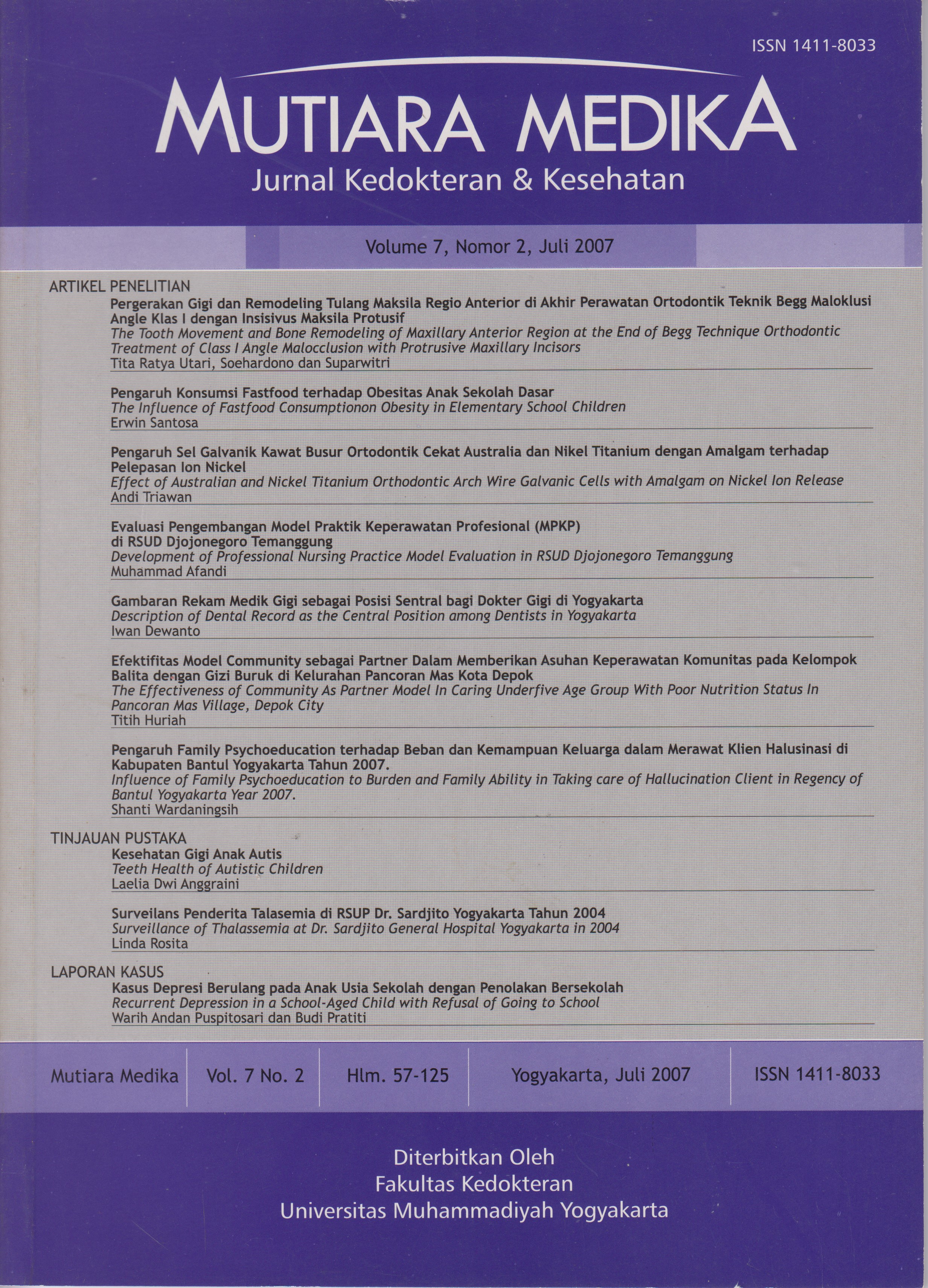
Pergerakan Gigi dan Remodeling Tulang Maksila Regio Anterior di Akhir Perawatan Ortodontik Teknik Begg Maloklusi Angle Klas I dengan Insisivus Maksila Protusif : Penelitian Deskriptif Observasional
DOI:
https://doi.org/10.18196/mmjkk.v7i2.1668Keywords:
rasio BT, sefalogram, teknik Begg tahap III, BT ratio, cephalogram, third stage Begg techniqueAbstract
Basic axiom in the orthodontic treatment is “bone trace tooth movement”, which means that a good orthodontic tooth movement is a tooth movement followed by remodeling of alveolus bone in the same degree (BT ratio 1:1). This study described tooth movement and bone remodeling of maxillary anterior region in the end of stage IIIBegg technique orthodontic treatment of Class I Angle malocclusion with protrusive maxillary incisors in adult patients who were treated in Orthodontic Clinic, Faculty of Dentistry, GadjahMada University from 1997 to 2005. Twenty-two cases fulfilled the criteria of research subjects. Cephalogram in the end of treatment was superimposed on the cephalogram of initial treatment for each case. The changes of point A described the amount of labial cortex bone remodeling, whereas the changes of maxillary incisor apex described the amount of tooth movement. The average of point A changes was 1.37 mm and average of incisors apex changes was 2.65 mm (ratio 1:1.93). The results demostrated that there was tooth movement which was greater than bone remodeling.
Aksioma dasar perawatan ortodontik adalah “bone trace tooth movement”, artinya pergerakan gigi ortodontik yang baik adalah pergerakan gigi yang diikuti remodeling tulang alveolus dengan derajat yang sama besar (Rasio BT 1:1). Penelitian ini mendiskripsikan pergerakan gigi dan remodeling tulang maksila regio anterior di akhir tahap III perawatan ortodontik teknik Begg maloklusi Angle klas I dengan insisivus maksila protusifpada pasien dewasa yang dirawat di Klinik Ortodonsia, Fakultas Kedokteran Gigi, Universitas Gadjah Mada dari tahun 1997 sampai 2005. Duapuluh dua kasus memenuhi syarat sebagai subyek penelitian. Sefalogram akhir perawatan disuperposisikan pada sefalogram awal perawatan dari setiap kasus. Perubahan titik A mendiskripsikan besarnya remodeling tulang korteks labial, sedangkan perubahan apeks insisivus maksila mendiskripsikan besarnya pergerakan gigi. Rerata perubahan titik A adalah 1,37 mm dan rerata perubahan apeks insisivus maksila adalah 2,65 mm (rasio 1:1,93). Hasil ini menunjukkan bahwa terjadi pergerakan gigi yang lebih besar dari pada remodeling tulangnya.
References
Cangialosi, TJ. and Meistrell, M.E., 1982, A cephalometric evaluation of hard- and soft-tissue changes during the third stage of Begg treatment, Am. J. Orthod., 81:124-129
De Angelis V, 1970, Observation on the response of alveolar bone to orthodontic force, Am. J. Orthod, 58:284-94
Gianelly A., and Goldman, H. M., 1971, Biologic Basis of Orthodontics, Lea & Febiger, Philadelphia., 116-38
Foster, T. D., 1993, Buku Ajar Ortodonsi, Edisi III, Penerbit Buku Kedokteran EGC, Jakarta, 168-85
Reitan, K., 1964, Effects of force magnitude and direction of tooth movement on different alveolar bone type, Angle Orthod, 34:244-55
Vardimon, A.D., Oren, E., and Ben- Bassat, Y, 1998, Cortical bone remodelling/tooth movement ratio during maxillary incisor retraction with tip versus torque movements, Am. J. Orthod. Dentofacial Orthop., 114:520¬529
Sarikaya, S., Haydar, B., Ciger, S., and Ariyurek, M., 2002, Change in alveolar bone thickness due to retraction of anterior teeth, Am. J. Orthod. Dentofacial Orthop., 122:15-26
Barrer, H.G., Buchin, I.D., Fogel, M.S., Swain, B.F., and Ackerman, J.L., 1971, Borderline extraction cases: Panel discussion. Part 5, J. Clin. Orthod., 5:609-26
Farrow, A.L., Zarinna, K., and Azizi, K., 1993, Bimaxillary protrusion in black Americans—an esthetic evaluation and treatment considerations, Am. J. Orthod. Dentofacial Orthop., 104:240¬50
Cadman, CAR, 1975, Avademecum for the Begg Technique: Technical Principal, Am. J. Orthod.,67(5):439-512
Begg, P.R., and Kesling, P.C., 1977/a, The differential force method of orthodontic treatment, Am. J. Orthod., 71(1):1-39
Kusnoto, H., 1977, Penggunaan Cephalometri Radiografi dalam Bidang Ortodonti, Bagian Orthodonti FKG Universitas Trisakti, Jakarta, 3-15
Jacobson, A. and Sadowsky, P. L., 1995, Superimposition of Cephalometric Radiographs, in Alexander Jacobson (ed) Radiography Cephalometric from Basics to Videoimaging, Quintessence Publishing Co. Inc.,Chicago
Begg, P.R., and Kesling, P.C., 1977/b, Begg Orthodontic Theory and Technique, 3 ed., W.B., Saunders Company Philadelphia, 192-93.
Downloads
Published
Issue
Section
License
Copyright
Authors retain copyright and grant Mutiara Medika: Jurnal Kedokteran dan Kesehatan (MMJKK) the right of first publication with the work simultaneously licensed under an Attribution 4.0 International (CC BY 4.0) that allows others to remix, adapt and build upon the work with an acknowledgment of the work's authorship and of the initial publication in Mutiara Medika: Jurnal Kedokteran dan Kesehatan (MMJKK).
Authors are permitted to copy and redistribute the journal's published version of the work (e.g., post it to an institutional repository or publish it in a book), with an acknowledgment of its initial publication in Mutiara Medika: Jurnal Kedokteran dan Kesehatan (MMJKK).
License
Articles published in the Mutiara Medika: Jurnal Kedokteran dan Kesehatan (MMJKK) are licensed under an Attribution 4.0 International (CC BY 4.0) license. You are free to:
- Share — copy and redistribute the material in any medium or format.
- Adapt — remix, transform, and build upon the material for any purpose, even commercially.
This license is acceptable for Free Cultural Works. The licensor cannot revoke these freedoms as long as you follow the license terms. Under the following terms:
Attribution — You must give appropriate credit, provide a link to the license, and indicate if changes were made. You may do so in any reasonable manner, but not in any way that suggests the licensor endorses you or your use.
- No additional restrictions — You may not apply legal terms or technological measures that legally restrict others from doing anything the license permits.


