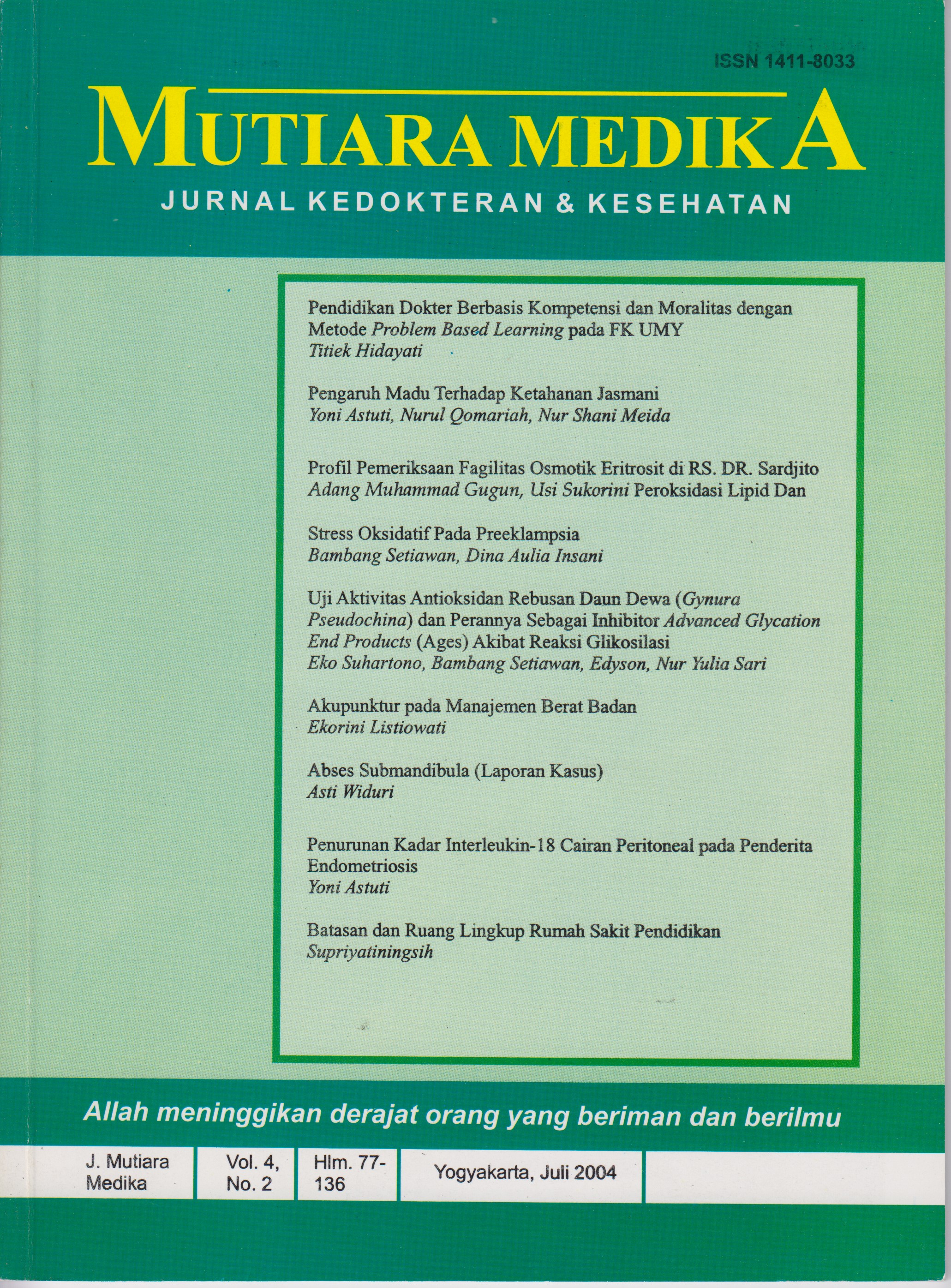
Profil Pemeriksaan Fragilitas Osmotik Eritrosit di RS. Dr. Sardjito
DOI:
https://doi.org/10.18196/mmjkk.v4i2.1745Keywords:
Test Osmotic Fragility, Thalassemia, hemolytic anemia, Osmotic Fragility testAbstract
Osmotic fragility test (OFT) is performed to differentiate anemia diagnose with erythrocyte physical changing. In thalassemia and hemolytic anemia, OFT probably gave variation results that can cause erroneous anemia etiology trac¬ing. Aims of this research are to describe the OFT profile and its comparison with peripheral blood morphology in thalassemia and hemolytic anemia.The method, this retrospective study was conducted in Dr. Sardjito hospital at January 2002 to June 2004. Chi-Square test was used to compare thalassemia and hemolytic anemia proportion in the OFT groups. OFT results from 61 sub¬jects were : increasing 17 (27,8%), increasing-decreasing 17 (27,8%), de¬creasing 15 (24,4%), and normal 12 (20%). There were significantly differ-ence proportions in thalassemia group between decreasing OFT to increasing and normal OFT (p-0,005 ; p=0,002), but no difference to increasing-de¬creasing group. In hemolytic anemia group, the difference proportion found significantly between increasing OFT to normal, increasing-decreasing and decreasing OFT (p=0,03; p-0,005; p=0,000, respectively). In increasing-de¬creasing OFT group, there was no difference in type anemia (p=0,32). Mor¬phologically, target cell was found in 81 % of thalassemia, and spherocyte in 70% of hemolytic anemia. In Dr. Sardjito Hospital, OFT gave variation profile and in Thalassemia and hemolytic anemia groups, morphology evaluation are needed to confirm OFT results.
Latar Belakang: Pemeriksaan fragilitas osmotik eritrosit (FOE) ini dilaksanakan untuk membantu diagnosis banding beberapa jenis anemia dengan sifat fisik eritrosit berubah. Aplikasi klinis, Talasemia dan anemia hemolitik memberikan hasil bervariasi sehingga dapat menimbulkan kesalahan interpretasi dalam melacak jenis maupun etiologi anemia.Tujuan penelitian ini adalah mengetahui variasi hasil FOE dan kesesuaian gambaran morfologi darah tepi pada talasemia dan anemia hemolitik. Penelitian retrospektif ini dilakukan menggunakan data rekam medik. Subyek adalah pasien yang diperiksa fragilitas osmotik eritrositnya di laboratorium Patologi Klinik RS. Dr. Sardjito antara Januari tahun 2002 sampai dengan Juni 2004. Uji Chi- square terhadap proporsi talasemia dan anemia hemolitik pada kelompok hasil FOE. Dari 61 subyek, variasi hasil FOE meliputi : peningkatan fragilitas 17 (27,8%), penurunan fragilitas 17 (24,4%), campuran peningkatan dan penurunan 15 (27,8%) dan normal 12 (20%). Terdapat perbedaan bermakna proporsi talasemia kelompok penurunun FOE terhadap kelompok peningkatan FOE (p=0,005) dan FOE normal (p= 0,002), namun tidak berbeda bermakna dengan hasil campuran penurunan dan peningkatan fragilitas (p= 0,26). Terdapat perbedaan bermakna proporsi ane¬mia hemolitik pada kelompok dengan peningkatan FOE terhadap kelompok normal FOE, campuran penurunan dan peningkatan FOE dan penurunan FOE (p =0,03; p= 0,005; p= 0,000). Tidak terdapat perbedaan bermakna proporsi jenis anemia pada hasil campuran penurunan dan peningkatan FOE (p= 0,32). Gambaran morfologi darah tepi pada kelompok talasemia, 81% memiliki sel target dan pada kelompok anemia hemolitik, 70% memiliki sel spherosit.Hasil FOE di RS Dr. Sardjito menunjukkan gambaran variasi, talasemia maupun anemia hemolitik membutuhkan konfirmasi morfologi darah tepi untuk meninjau kesesuaiannya.
References
Stiene- Martin EA, Lotspeich-Steininger CA, Koepke JA, 1998. Clinical Haematology Principles, Procedures, Correlatoins, 2nd ed. Lippincot Philadelphia, New York, 193-265.
Kjeldsberg CR, 1995. Practical Diagnosis of Hematologic Disorders, 2th ed American Society of Clinical Pathologists, Chicago Illinois, 109-162
Brown BA, 1993. Hematology Principles and Procedures, 6* ed. Lippincot Williams and Walkins, Philadelphia, 174-180.
Hoffbrand AU. Pettit JE, 1992. Kapita Selekta Hematologi, EGC, Jakarta.
Dacie JV.,Lewis SM, 1991. Practical Haematology, 7th ed. Churchill Livingstone Edinburgh Lon¬don, Melbourne and New York, 175-256.
Anonim, 2004. Prosedur Tetap Pemeriksaan Sub Hematologi, Instalasi Patologi Klinik RS. Dr. Sardjito.
Ronald A. Sacher, Richard A. Mc Pherson, 2004 Tinjauan Klinis Hasil Pemeriksaan Laboratorium , Edisi 11 EGC Jakarta, 82-108.
Suphan Soogarun, Viroj Wiwanitkit, Jamsai Suwansaksri, Nara Paritpokee, 2004. The Prevalence of Fragile Red Cell for Talasemia, Department of Clinical Microscopy, Faculty of Allied Health Science, Chulalongkom University, Bangkok, Thailand.
E. Setoudeh Maram, Mohtasram Amiri, M. Haghshenas 2000. Effectiveness of Osmotic Fragility Screening with varying saline Concentration in Detecting a- Talasemia Trait. Iran Jomal Medicine Science 25 (1&2), 56-58.
Sanjay Kumar, 2002. An Analogy for Explaining Erythrocyte Fragility :Concepts made easy, Advances in Phisiology Education - June.
Giger Urs , 2000. Regenerative Anemias Caused by Blood Loss of hemolysis, “Textbook of Veterinary Internal Medicine, S.J. Ettinger an E.C. Felrnan, ed. Philadelphia, PA Saunders.
Elghetany and Davey FR,1996. Erythrocyte Disorders. In Clinical Diagnosis and managemeny by Laboratory Methods, 19 ed. JB Henry, ed, Philadelphia: WB Saunders Co, 634.
Gladen BE, Luicens JN, 1999. Hereditary Spherocytosis and Other Anemias oe to Abnormalities of the Red Cell Membrane. In Wintrobe’s Clinical Hematology. 10th ed. Baltimore, William and Wilkins, p 1132-1159.
Gallagher PG, Jarolin P, 2000. Red cell membrane disorder. In Hematology: Basic Principles and Practise. 3rd New York Churchill Livingstone, p 576-610.
Mazeron et al. Theoretical Approach of The Measurement of Osmotic Fragility of Erythrocytes by Optical transmissions . Journal: Photochemistry and Photobilogy Volume 72 Issue: 2 Pages: 172-178.
Downloads
Published
Issue
Section
License
Copyright
Authors retain copyright and grant Mutiara Medika: Jurnal Kedokteran dan Kesehatan (MMJKK) the right of first publication with the work simultaneously licensed under an Attribution 4.0 International (CC BY 4.0) that allows others to remix, adapt and build upon the work with an acknowledgment of the work's authorship and of the initial publication in Mutiara Medika: Jurnal Kedokteran dan Kesehatan (MMJKK).
Authors are permitted to copy and redistribute the journal's published version of the work (e.g., post it to an institutional repository or publish it in a book), with an acknowledgment of its initial publication in Mutiara Medika: Jurnal Kedokteran dan Kesehatan (MMJKK).
License
Articles published in the Mutiara Medika: Jurnal Kedokteran dan Kesehatan (MMJKK) are licensed under an Attribution 4.0 International (CC BY 4.0) license. You are free to:
- Share — copy and redistribute the material in any medium or format.
- Adapt — remix, transform, and build upon the material for any purpose, even commercially.
This license is acceptable for Free Cultural Works. The licensor cannot revoke these freedoms as long as you follow the license terms. Under the following terms:
Attribution — You must give appropriate credit, provide a link to the license, and indicate if changes were made. You may do so in any reasonable manner, but not in any way that suggests the licensor endorses you or your use.
- No additional restrictions — You may not apply legal terms or technological measures that legally restrict others from doing anything the license permits.


