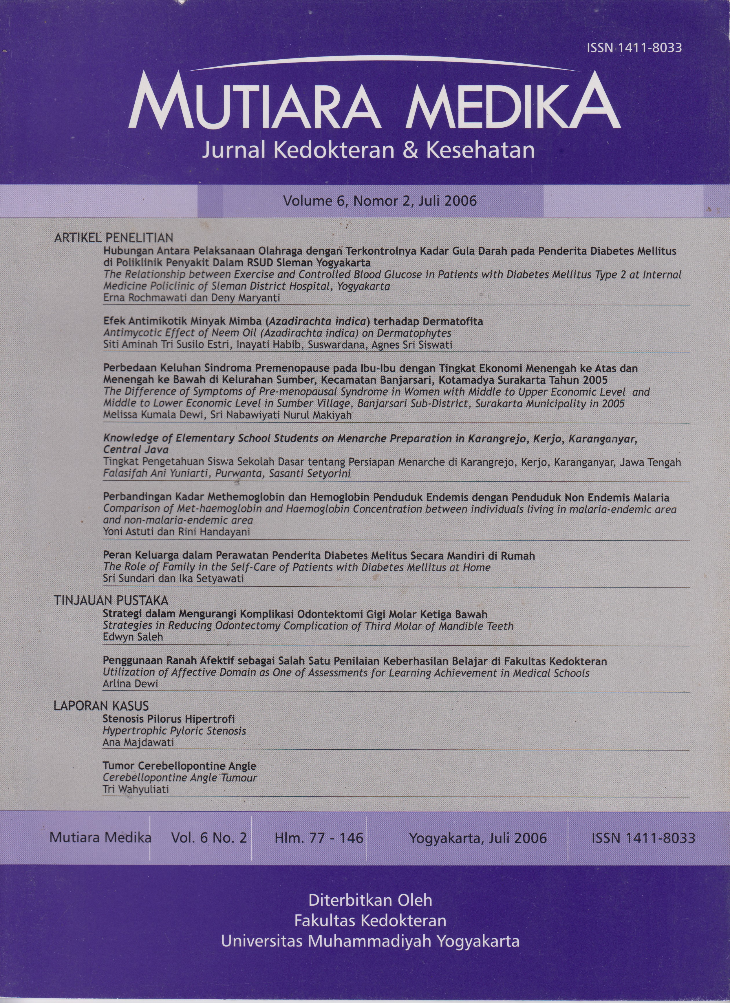
Tumor Cerebellopontine Angle
DOI:
https://doi.org/10.18196/mmjkk.v6i2.1897Abstract
For about 85% of tumors in the region of the cerebellopontine angle is acoustic neuroma, 10% are meningiomas and 5% are epidermoid tumor. An acoustic neuroma is a benign tumour that develops in the acoustic or auditory nerve. The acoustic neuroma starts from schwann cells which cover the acoustic nerve and is sometimes therefore called a schwannoma.Hearing loss is the most frequent symptom. Vertigo or spinning occurs in about 20 percent of persons with acoustic neuroma. Early diagnose by signs and symptoms detection is very important in acoustic neuroma treatment. Unfortunately, misdiagnose becomes the problem in this step. The optimal test for excluding an acoustic neuroma is MRI. If an MRI cannot be done, CT scan should be obtained.
About more than a half of all acoustic neuromas are presently treated with surgery. The recurrence rate of the tumor is about 3% after surgery. Overall, the risk of death from acoustic neuroma surgery is about 0.5 to 2 percent. Unexpected post-operative complications occur in roughly 20percent with more complications occurring in elderly and infirm individuals and those with large tumors. Surgical treatment where the brain is exposed is nearly always performed by a team of surgeons, usually including a neurologist, a neurosurgeon and an anesthesiolgist. Pathology anatomist is needed to make definitif diagnose.
Sekitar 85% tumor yang tumbuh di daerah sudut serebelo pontin adalah neuroma akustik, sekitar 10% adalah meningioma dan sekitar 5% adalah tumor epidermoid. Neuroma akustik adalah tumor jinak yang tumbuh pada nervus akustikus, yang berasal dari sel schwann yang meliputi nervus akustikus sehingga disebut juga schwanoma.
Gangguan pendengaran merupakan gejala yang paling sering ditemukan. Vertigo atau rasa berputar dirasakan pada sekitar 20% penderita. Diagnosa dini melalui pengenalan tanda dan gejala sangat penting dalam penatalaksanaan neuroma akustik. Sayangnya, hal ini seringkah terabaikan atau teijadi misdiagnosis. Pemeriksaan yang optimal adalah menggunakan MRI, namun jika hal itu tidak bisa dilakukan maka CT scan sebaiknya dilakukan.
Sekitar lebih dari setengah kasus neuroma akustik diterapi melalui pembedahan. Angka kekambuhan setelah pembedahan sekitar 3%. Risiko kematian saat operasi sekitar 0,5 - 2%. Komplikasi setelah operasi muncul pada sekitar 20% penderita, yang paling banyak pada penderita usia dewasa serta pada tumor yang besar. Terapi pembedahan pada otak melibatkan tim yang terdiri dari dokter ahli saraf, bedah saraf dan ahli anestesi. Ahli patologi anatomi berperan dalam menentukan diagnosa pasti.
References
Greenberg.MS. Handbook of Neurosurgery, Fifth ed., thieme medical Publisher, Greenberg Graphics Inc., new York. 2001.
Duus P. Diagnosis Topik Neurologi: anatomi, fisiologi, tanda, gejala. Ed.2 , 1996.
Grace F Kao. 2004. Neurilemoma. Emedicine, Last Updated: January 20, 2004.
Satyanegara. Ilmu bedah saraf; editor, L. Djoko Listiono.-Ed 3,1998
Gilroy. Basic Neurology, 2nd ed.Mc Graw Hil Inc. Pergamon Pres. Singapore, 1992
Lindsay, K., Bone, I., Callander, R., 1997 Neurology and Neurosurgery Illustrated, 3th ed. Churchill, Livingstone, Tokyo
Pothula VB,Lesser T, Mallucci C, may P, Foy P, Vestibular schwannomas in children. Otol Neurotol 22: 903-907, 2001
Timothy C.Heen MD, Acoustic Neuroma, April 2004
Downloads
Issue
Section
License
Copyright
Authors retain copyright and grant Mutiara Medika: Jurnal Kedokteran dan Kesehatan (MMJKK) the right of first publication with the work simultaneously licensed under an Attribution 4.0 International (CC BY 4.0) that allows others to remix, adapt and build upon the work with an acknowledgment of the work's authorship and of the initial publication in Mutiara Medika: Jurnal Kedokteran dan Kesehatan (MMJKK).
Authors are permitted to copy and redistribute the journal's published version of the work (e.g., post it to an institutional repository or publish it in a book), with an acknowledgment of its initial publication in Mutiara Medika: Jurnal Kedokteran dan Kesehatan (MMJKK).
License
Articles published in the Mutiara Medika: Jurnal Kedokteran dan Kesehatan (MMJKK) are licensed under an Attribution 4.0 International (CC BY 4.0) license. You are free to:
- Share — copy and redistribute the material in any medium or format.
- Adapt — remix, transform, and build upon the material for any purpose, even commercially.
This license is acceptable for Free Cultural Works. The licensor cannot revoke these freedoms as long as you follow the license terms. Under the following terms:
Attribution — You must give appropriate credit, provide a link to the license, and indicate if changes were made. You may do so in any reasonable manner, but not in any way that suggests the licensor endorses you or your use.
- No additional restrictions — You may not apply legal terms or technological measures that legally restrict others from doing anything the license permits.


