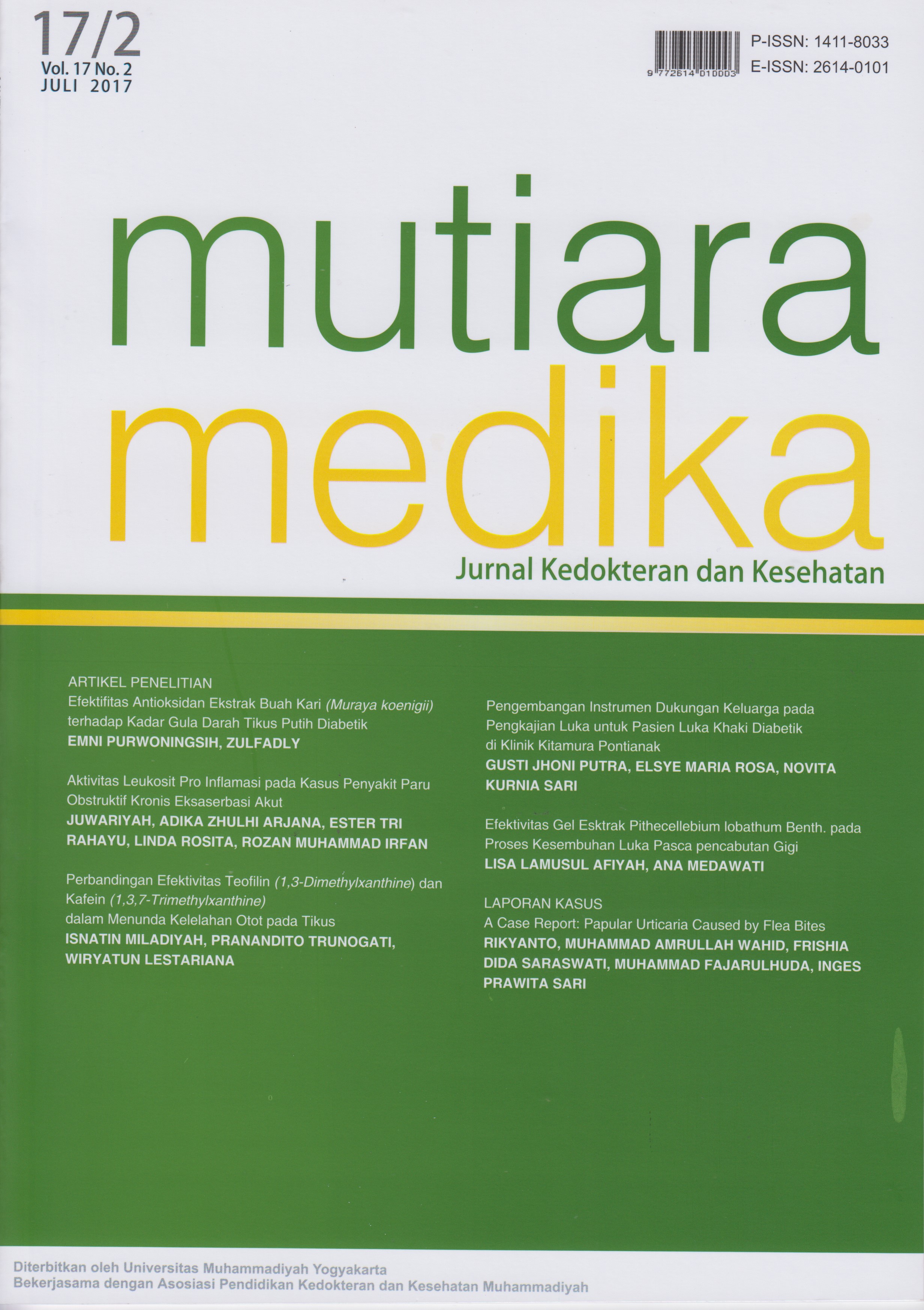
Aktivitas Leukosit Pro Inflamasi pada Kasus Penyakit Paru Obstruktif Kronis Eksaserbasi Akut
DOI:
https://doi.org/10.18196/mm.170202Keywords:
Eosinofil, Netrofil, PPOKAbstract
Penyakit Paru Obstruktif Kronis (PPOK) memiliki karateristik adanya restriksi saluran nafas yang kurang reversibel. Pada PPOK terdapat inflamasi akibat aktifitas sel-sel inflamasi termasuk neutrofil dan eosinofil. Restriksi saluran nafas terjadi akibat remodelling dari proses inflamasi yang terjadi. Penelitian ini bertujuan untuk mengetahui aktifitas sel-sel inflamasi terutama neutrofil dan eosinofil pada PPOK eksaserbasi akut dengan membandingkan kadar eosinfil dan netrofil sebelum dan sesudah terapi. Penelitian bersifat observasional dengan desain cross sectional. Responden penelitian ini adalah pasien penderita PPOK yang rawat jalan dan rawat inap di RSUD Kebumen pada tahun 2016. Semua subyek masuk dalam penelitian dengan kriteria eksklusi adalah data tidak lengkap. Hasil menunjukkan terdapat 119 pasien yang memenuhi kriteria inklusi sebagai responden. Data dari rekam medis menunjukkan bahwa mayoritas penderita adalah laki-laki (84,03 %) dengan rata-rata umur 67 tahun. Penyakit penyerta yang ditemukan adalah hipertensi (54,62 %), tuberkulosis (22,69 %) dan congestive heart failure (CHF) (6,72 %). Pada tanda vital, terdapat kenaikan sistole dan laju nafas. Presentase netrofil pada kedua jenis kelamin meningkat dibandingkan normal namun tidak dengan eosinofil. Setelah dilakukan rawat inap, terjadi penurunan persentase eosinofil dan neutrofil dibanding sebelum perawatan namun tidak signifikan secara statistik (p= 0,603 vs 0,818). Kesimpulan dari penelitian ini adalah adanya peningkatan aktivitas netrofil pada pasien PPOK. Penurunan aktivitas baik netrofil maupun eosinofil didapatkan ketika pasien rawat inap meskipun tidak bermakna secara statistik.
References
Kasper DL, Fauci AS, Longo DL, Braunwald E, Hauser SL, Jameson JL. Harrison’s Principles of Internal Medicine. 16th ed. New York: The McGraw-Hill Companies. 2005.
Gorczynski R, Stanley J. Clinical immunology. Texas: landes Bioscience. 1999.
Cruse JM, Lewis RE. Illustrated Dictionary of Immunology (2nd Edition). London, UK: Taylor & Francis. 2003.
Hoppenot D, Malakauskas K, Lavinskienė S, Bajoriūnienė I, Kalinauskaitė V, Sakalauskas R. Peripheral Blood Th9 Cells and Eosinophil Apoptosis in Asthma Patients. Medicina (Kaunas), 2015; 51 (1): 10–17.
Saeki M, Kaminuma O, Nishimura T, Kitamura N, Mori A, Hiroi T. Th9 Cells Elicit EosinophilIndependent Bronchial Hyperresponsiveness in Mice. Allergol Int, 2016; 65 (Suppl): S24–S29.
Lukawska JJ, Livieratos L, Sawyer BM, Lee T, O’Doherty M, Blower PJ, et al. Imaging Inflammation in Asthma: Real Time, Differential Tracking of Human Neutrophil and Eosinophil Migration in Allergen Challenged, Atopic Asthmatics in Vivo. EBioMedicine, 2014; 1 (2): 173–180.
Mazzeo C, Cañas JA, Zafra MP, Rojas MA, Fernández-Nieto M, Sanz V, et al. Exosome Secretion by Eosinophils: A Possible Role in Asthma Pathogenesis. J Allergy Clin Immunol, 2015; 135 (6): 1603–1613.
Zeiger RS, Schatz M, Dalal AA, Chen W, Sadikova E, Suruki RY, et al. Blood Eosinophil Count and Outcomes in Severe Uncontrolled Asthma: A Prospective Study. J Allergy Clin Immunol Pract, 2016; 5 (1): 144-153.
Karch A, Vogelmeier C, Welte T, Bals R, Kauczor HU, Biederer J, et al. The German COPD Cohort COSYCONET: Aims, Methods and Descriptive Analysis of the Study Population at Baseline. Respir Med. 2016; 114 (1): 27–37.
Hagstad S, Backman H, Bjerg A, Ekerljung L, Ye X, Hedman L, et al. Prevalence and Risk Factors of COPD among Never-Smokers in Two Areas of Sweden - Occupational Exposure to Gas, Dust or Fumes is an Important Risk Factor. Respir Med, 2015; 109 (11): 1439–1445.
Jenkins CR, Chapman KR, Donohue JF, Roche N, Tsiligianni I, Han MK. Improving the Management of COPD in Women. Chest. 2016; 151 (3): 686-696.
Melzer AC, Ghassemieh BJ, Gillespie SE, Lindenauer PK, McBurnie MA, Mularski RA, et al. Patient Characteristics Associated with Poor Inhaler Technique among a Cohort of Patients with COPD. Respir Med, 2017; 123 (1): 124-130.
Fragoso E, Andr S, Boleo-Tom JP, Areias V, Munh J, Cardoso J. Understanding COPD: A Vision on Phenotypes, Comorbidities and Treatment Approach. Rev Port Pneumol. 2016; 22 (2): 101–111.
Global Initiative for Chronic Obstructive Lung Disease. Global Strategy for the Diagnosis, Management and Prevention of Chronic Obstructive Pulmonary Disease. 2006.
O’Toole RF, Shukla SD, Walters EH. TB Meets COPD: An Emerging Global Co-Morbidity in Human Lung Disease. Tuberculosis. 2015; 95 (6): 659–663.
Gunen H, Yakar H. The Role of TB in COPD. Chest, 2016; 150 (4): 856A.
Mohamed-Hussein AAR, Gamal EW, Abd Allah MS. Value of Blood Eosinophilia in PhenotypeDirected Corticosteroid Therapy of COPD Exacerbation: Final Results. Egypt J Chest Dis Tuberc, 2016; 66 (1): 221-225.
Pedersen F, Marwitz S, Holz O, Kirsten A, Bahmer T, Waschki B, et al. Neutrophil Extracellular Trap Formation and Extracellular DNA in Sputum of Stable COPD Patients. Respir Med, 2015; 109 (10): 1360–1362.
Yousef AM, Alkhiary W. Role of Neutrophil to Lymphocyte Ratio in Prediction of Acute Exacerbation of Chronic Obstructive Pulmonary Disease. Egypt J Chest Dis Tuberc, 2016; 66 (1): 1-6.
Kuna P, Jenkins M, O’Brien CD, Fahy WA. AZD. A Neutrophil Elastase Inhibitor, Plus ongoing Budesonide/ Formoterol in Patients with COPD. Respir Med, 2012; 106 (4): 531–539.
Gupta V, Khan A, Higham A, Lemon J, Sriskantharajah S, Amour A, et al. The Effect of Phosphatidylinositol-3 Kinase Inhibition on Matrix Metalloproteinase-9 and Reactive Oxygen Species Release from Chronic Obstructive Pulmonary Disease Neutrophils. Int Immunopharmacol, 2016; 35 (1): 155–162.
Oudijk EJD, Gerritsen WBM, Nijhuis EHJ, Kanters D, Maesen BLP, Lammers JWJ, et al. Expression of Priming-Associated Cellular Markers on Neutrophils during an Exacerbation of COPD. Respir Med, 2006; 100 (10): 1791– 1799.
Lerner CA, Sundar IK, Rahman I. Mitochondrial Redox System, Dynamics and Dysfunction in Lung Inflammaging and COPD. Int J Biochem Cell Biol, 2016; 81 (Pt. B): 294–306.
Downloads
Published
Issue
Section
License
Copyright
Authors retain copyright and grant Mutiara Medika: Jurnal Kedokteran dan Kesehatan (MMJKK) the right of first publication with the work simultaneously licensed under an Attribution 4.0 International (CC BY 4.0) that allows others to remix, adapt and build upon the work with an acknowledgment of the work's authorship and of the initial publication in Mutiara Medika: Jurnal Kedokteran dan Kesehatan (MMJKK).
Authors are permitted to copy and redistribute the journal's published version of the work (e.g., post it to an institutional repository or publish it in a book), with an acknowledgment of its initial publication in Mutiara Medika: Jurnal Kedokteran dan Kesehatan (MMJKK).
License
Articles published in the Mutiara Medika: Jurnal Kedokteran dan Kesehatan (MMJKK) are licensed under an Attribution 4.0 International (CC BY 4.0) license. You are free to:
- Share — copy and redistribute the material in any medium or format.
- Adapt — remix, transform, and build upon the material for any purpose, even commercially.
This license is acceptable for Free Cultural Works. The licensor cannot revoke these freedoms as long as you follow the license terms. Under the following terms:
Attribution — You must give appropriate credit, provide a link to the license, and indicate if changes were made. You may do so in any reasonable manner, but not in any way that suggests the licensor endorses you or your use.
- No additional restrictions — You may not apply legal terms or technological measures that legally restrict others from doing anything the license permits.


