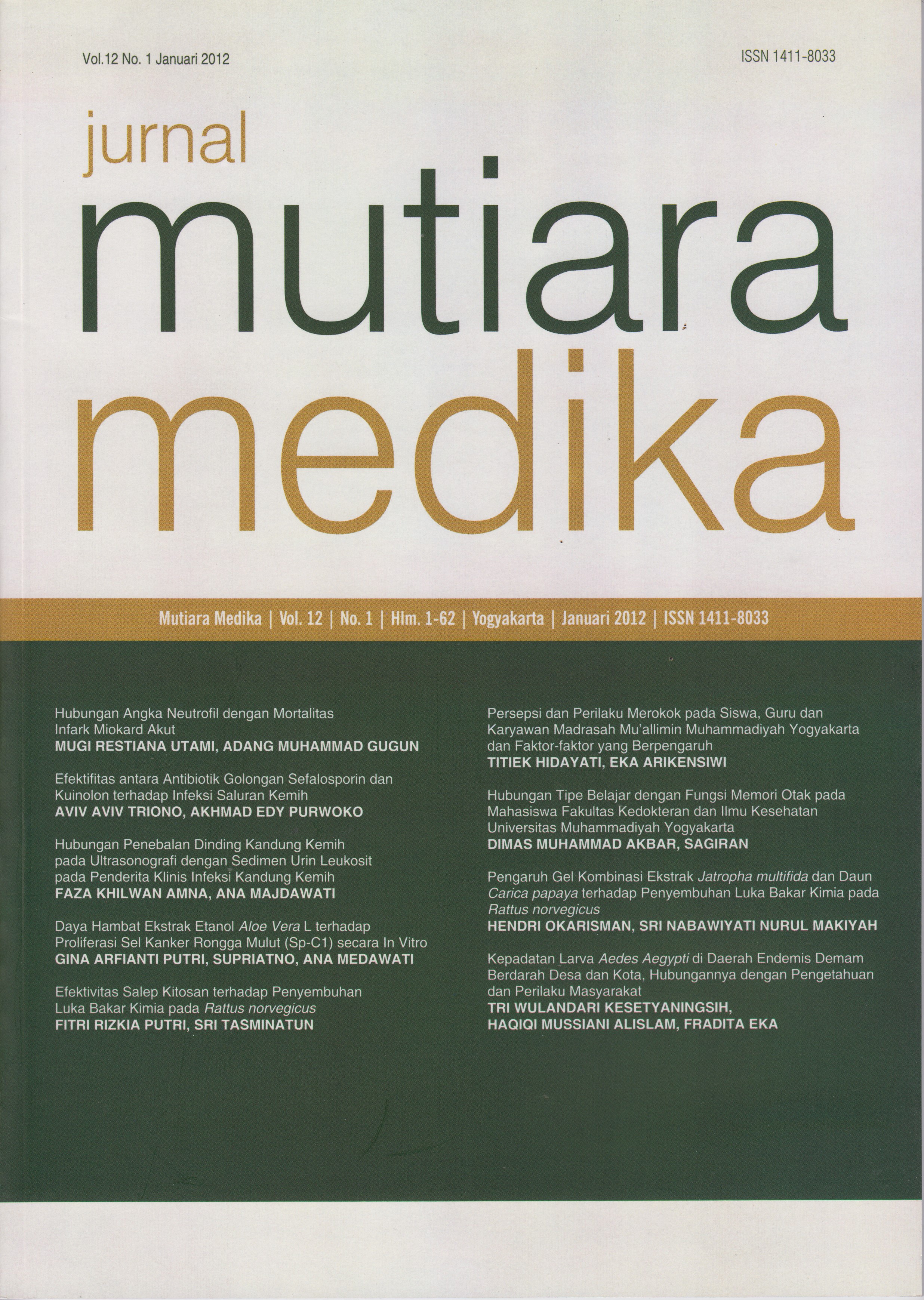
Hubungan Penebalan Dinding Kandung Kemih pada Ultrasonografi dengan Sedimen Urin Leukosit pada Penderita Klinis Infeksi Kandung Kemih
DOI:
https://doi.org/10.18196/mmjkk.v12i1.995Keywords:
Infeksi Saluran Kemih (ISK), Sistitis, Ultrasonografi, sedimen urin leukosit, Urinary Tract Infection (UTI), bladder infection, ultrasonography, urine sediment leukocytesAbstract
Infeksi Saluran Kemih (ISK) merupakan infeksi berupa pertumbuhan dan perkembangbiakan mikroorganisme dalam saluran kemih yang meliputi ginjal sampai kandung kemih,diantaranya Sistitis ( infeksi kandung kemih). Ultrasonografi (USG) dasawarsa terakhir ini merupakan pemeriksaan yang sering digunakan sebagai pilihan penunjang diagnostik pada beberapa kasus yang berhubungan dengan infeksi kandung kemih. Sedimen urin leukosit merupakan pemeriksaan semikuantitatif sebagai penunjang diagnosis Sistitis dengan acuan kadar sedimen urin leukosit positif. Penelitian ini bertujuan untuk mengetahui hubungan penebalan dinding kandung kemih pada pemeriksaan USG dengan sedimen urin leukosit pada penderita dengan klinis Sistitis. Desain penelitian ini observasional dengan studi cross sectional,menggunakan data sekunder dari catatan rekam medis pasien RS PKU Muhammadiyah I-II Yogyakarta untuk semua kasus ISK periode 1 Juli 2010 sampai 31 Agustus 2011. Data rekam medis yang digunakan adalah subyek penelitian dengan suspek infeksi kandung kemih yang mempunyai hasil laboratorium urin (sedimen urin lekosit) dan tebal dinding kandung kemih potongan transversal dan longitudinal pada pemeriksaan USG. Hasil analisis data dengan uji chi square didapatkan nilai p 0,631, sehingga dapat disimpulkan bahwa tidak terdapat hubungan penebalan dinding kandung kemih pada USG dengan hasil pemeriksaan sedimen urin leukosit.
Urinary Tract Infection (UTI) is a form of infection that involve growth and proliferation of microorganisms in the urinary tract includes kidney to the bladder, one type of UTI is cystitis (bladder infection). Ultrasonography (USG) examination in the last decade is frequently used as a diagnostic support in some cases associated with bladder infection. Examination of leukocyte urine sediment is a semiquantitative test that could be supporting a diagnosis of bladder infection with reference levels of urine sediment positive leukocytes. This study aims to determine the relationship between bladder wall thickening on ultrasonography with urine sediment of leukocytes in patients with clinical bladder infection. The study design was observational with cross sectional study using secondary data from the medical records of PKU Muhammadiyah Hospital of Yogyakarta I-II for all cases of Urinary Tract Infection in the period July 1, 2010 until August 31, 2011. Medical record data used in this study were research subjects with suspected bladder infection who had a urine laboratory results (urine sediment leucocytes) and bladder wall thickness transverse and longitudinal cuts on ultrasound examination. The results of data analysis with Chi-Square test p-value obtained 0.631. There is no relationship between the thickening of the bladder on ultrasonography with urine sediment leukocyte.
References
Kapur, J., & Stringer, D.A. Diagnostic Imaging and Intervention: A Guide for Clinicians. In Chiu MC, Yap HK (eds). Practical Paediatric Nephrology : an Update of Current Practice. Medcom Limited, Hong Kong. 2005. p.15-29
Aiyegoro, O.A., Igbinosa, O.O., Ogunmwonyi, I.N., Odjadjare, E.E., Igbinosa, O.E., Okoh, A. Incidence of urinary tract infections (UTI) among children and adolescents in Ile-Ife Nigeria. Afr. J. Microbiol. Res, 2007 july: 013-019.
Agus, T. Infeksi Saluran Kemih, Buku Ajar Ilmu Penyakit Dalam Jilid I Edisi IV. Jakarta: FK UI. 2001.
Santosa, A., Tjahjodjati, R.A., Tarmono, A. Panduan Penatalaksanaan (Guidelines) Pediatric Urology (Urologi Anak) di Indonesia; Infeksi Saluran Kemih. 2005. Diakses tanggal 5 April 2011 dari http://www.google.com/url?sa=t&rct = j&q=Panduan+Penatalaksanaan+%28Guidelines% 29+ Pediatric+Urology+%28Urologi+Anak%29+di + Indonesia%3B++Infeksi+Saluran+Kemih&source= web&cd=1&cad=rja&ved=0CCgQFjAA&url=http%3 A%2F%2Fwww.iaui.or.id%2Fast%2Ffile%2Fpediatric_ urology.doc&ei=9sHwUfHNDsnprQf8yoCoDw &usg=AFQjCNERfX0M5KAZVBpCWT9 WWT NyNe8avw
Wirawan, R., Astrawinata, D. A. W., Enny. Evaluasi pemeriksaan sedimen urin secara kuantitatif menggunakan sistem Shih-Yung. Jakarta: Bagian Patologi Klinik, FK UI/RSUPN Dr. Ciptomangunkusomo. 2004.
Kelly, CE. The relationship between pressure flow studies and ultrasound estimates bladder wall mass. Urology Journal, 2005; 7 (6): S29S34.
Jequier S. & Rousseau O. 1987. Sonographic measurements of the normal bladder wall in children. AJR Am J Roentgenol. 1987; 149 (3): 563-6.
Yang JM, Huang WC. Bladder Wall thickness on Ultrasonographic cystourethrography: Affecting factors and their implications. J Ultrasound Med. 2003; 22 (8): 777-82.
Wald, R., Bell, C.M., Neisenbaum, R., Perrone, S., Liangos, O., Laupacis, A., et al. Interobsever Reliability of Urin Sediment Interpretation. Clin J Am Soc Nephrol, 2009; 4 (3): 567-571. 10. De Jong, W. Buku Ajar Ilmu Bedah, edisi 2. Jakarta: EGC. 2004.
Shortliffe & McCue J.D. Urinary Tract Infection at The Age Extremes: Pediatric and Geriatric (Abstract). Am J Med, 2002; 113 (Suppl 1A): 55S-66S.
Brunzel, N.A. Fundamentals of Urine and Body Fluid Analysis. Philadelphia: WB Saunders Company, 2004. p. 119-263.
Sorkhi, R.M., Nooreddini, H.G., Navase, A.R., Shafee, H., Hadipoor, A. Sonographic measurement of bladder wall thickness in healthy children. Iranian Journal of Pediatrics, 2009 ; 19 (4): 341-6.
Lim, R. 2009. Vesicoureteral Reflux and Urinary Tract Infection: Evolving Practices and Current Controversies In Pediatric Imaging. AJR Am J Roentgenol, 2009; 192 (5): 11971208.
Rosita, L. Pengaruh Penundaan Waktu Terhadap Hasil Urinalisis. Yogyakarta: Departemen Patologi Klinik, Fakultas Kedokteran Universitas Islam Indonesia. 2008.
Gandasoebrata, R. Urinalisis, dalam: Penuntun Laboratorium Klinik, cetakan ke-10. Jakarta: PT. Dian Rakyat; 2001. p. 112-263.
Aulia, D. Pemeriksaan dan Penilaian Kimia Urin dengan Carik Celup. Dalam: Kumpulan Makalah Lokakarya Aspek Praktis Urinalisis. Pendidikan Berkesinambungan Patologi Klinik. Jakarta: FK UI; 2004. p. 23-30.
Palmer, P.S. Panduan Pemeriksaan Diagnostik USG. WHO: EGC. 2002.
Downloads
Published
Issue
Section
License
Copyright
Authors retain copyright and grant Mutiara Medika: Jurnal Kedokteran dan Kesehatan (MMJKK) the right of first publication with the work simultaneously licensed under an Attribution 4.0 International (CC BY 4.0) that allows others to remix, adapt and build upon the work with an acknowledgment of the work's authorship and of the initial publication in Mutiara Medika: Jurnal Kedokteran dan Kesehatan (MMJKK).
Authors are permitted to copy and redistribute the journal's published version of the work (e.g., post it to an institutional repository or publish it in a book), with an acknowledgment of its initial publication in Mutiara Medika: Jurnal Kedokteran dan Kesehatan (MMJKK).
License
Articles published in the Mutiara Medika: Jurnal Kedokteran dan Kesehatan (MMJKK) are licensed under an Attribution 4.0 International (CC BY 4.0) license. You are free to:
- Share — copy and redistribute the material in any medium or format.
- Adapt — remix, transform, and build upon the material for any purpose, even commercially.
This license is acceptable for Free Cultural Works. The licensor cannot revoke these freedoms as long as you follow the license terms. Under the following terms:
Attribution — You must give appropriate credit, provide a link to the license, and indicate if changes were made. You may do so in any reasonable manner, but not in any way that suggests the licensor endorses you or your use.
- No additional restrictions — You may not apply legal terms or technological measures that legally restrict others from doing anything the license permits.


